• 文献检索
  • 文档翻译
  • 深度研究
  • 学术资讯
  • Suppr Zotero 插件Zotero 插件
  • 邀请有礼
  • 套餐&价格
  • 历史记录
应用&插件
Suppr Zotero 插件Zotero 插件浏览器插件Mac 客户端Windows 客户端微信小程序
定价
高级版会员购买积分包购买API积分包
服务
文献检索文档翻译深度研究API 文档MCP 服务
关于我们
关于 Suppr公司介绍联系我们用户协议隐私条款
关注我们

Suppr 超能文献

核心技术专利:CN118964589B侵权必究
粤ICP备2023148730 号-1Suppr @ 2026

文献检索

告别复杂PubMed语法,用中文像聊天一样搜索,搜遍4000万医学文献。AI智能推荐,让科研检索更轻松。

立即免费搜索

文件翻译

保留排版,准确专业,支持PDF/Word/PPT等文件格式,支持 12+语言互译。

免费翻译文档

深度研究

AI帮你快速写综述,25分钟生成高质量综述,智能提取关键信息,辅助科研写作。

立即免费体验

深圳技术大学-中山大学自动化渗出物检测和糖尿病性视网膜病变分级数据集。

The SUSTech-SYSU dataset for automated exudate detection and diabetic retinopathy grading.

机构信息

Department of Electrical and Electronic Engineering, Southern University of Science and Technology, Shenzhen, 5180000, China.

School of Electronics and Information Technology, Sun Yat-sen University, Guangzhou, 510000, China.

出版信息

Sci Data. 2020 Nov 20;7(1):409. doi: 10.1038/s41597-020-00755-0.

DOI:10.1038/s41597-020-00755-0
PMID:33219237
原文链接:https://pmc.ncbi.nlm.nih.gov/articles/PMC7679367/
Abstract

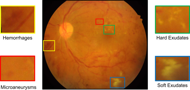
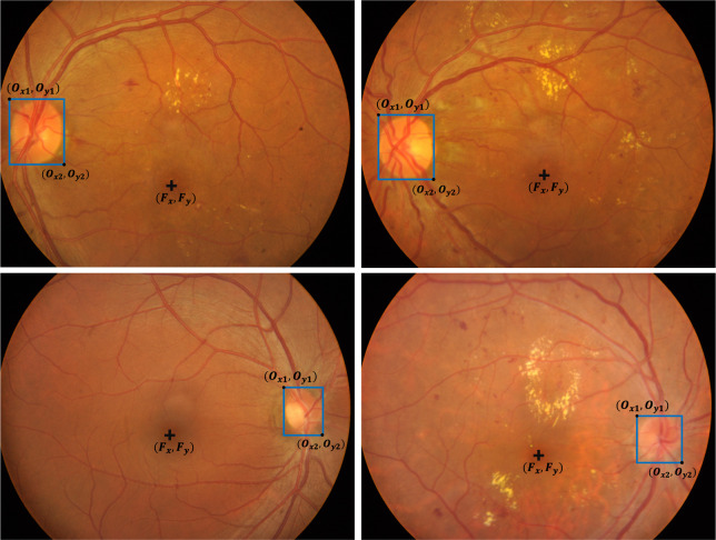
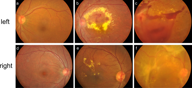
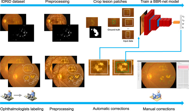
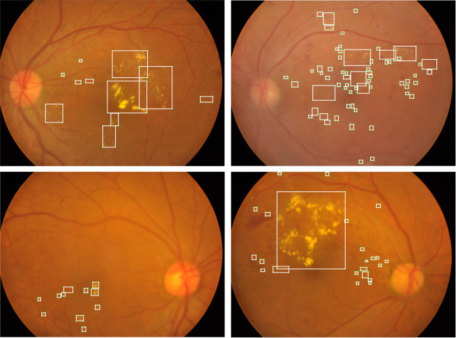
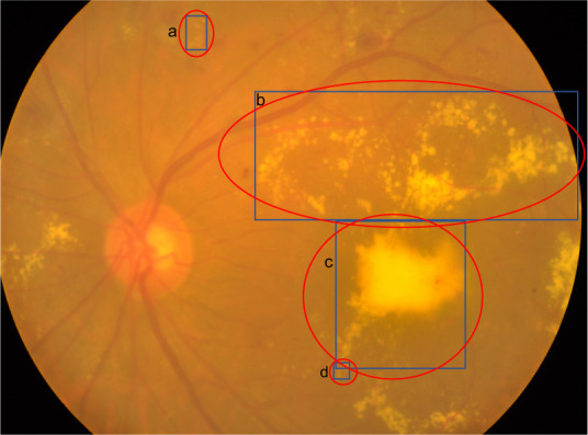
Automated detection of exudates from fundus images plays an important role in diabetic retinopathy (DR) screening and evaluation, for which supervised or semi-supervised learning methods are typically preferred. However, a potential limitation of supervised and semi-supervised learning based detection algorithms is that they depend substantially on the sample size of training data and the quality of annotations, which is the fundamental motivation of this work. In this study, we construct a dataset containing 1219 fundus images (from DR patients and healthy controls) with annotations of exudate lesions. In addition to exudate annotations, we also provide four additional labels for each image: left-versus-right eye label, DR grade (severity scale) from three different grading protocols, the bounding box of the optic disc (OD), and fovea location. This dataset provides a great opportunity to analyze the accuracy and reliability of different exudate detection, OD detection, fovea localization, and DR classification algorithms. Moreover, it will facilitate the development of such algorithms in the realm of supervised and semi-supervised learning.

摘要

眼底图像中渗出物的自动检测在糖尿病性视网膜病变(DR)筛查和评估中起着重要作用,为此通常首选监督式或半监督式学习方法。然而,基于监督式和半监督式学习的检测算法的一个潜在局限性是,它们在很大程度上依赖于训练数据的样本量和注释的质量,这正是这项工作的基本动机。在这项研究中,我们构建了一个包含 1219 张眼底图像(来自 DR 患者和健康对照者)的数据集,这些图像带有渗出病变的注释。除了渗出物的注释,我们还为每张图像提供了四个附加标签:左眼与右眼标签、来自三个不同分级方案的 DR 等级(严重程度量表)、视盘(OD)的边界框和黄斑位置。该数据集为分析不同渗出物检测、OD 检测、黄斑定位和 DR 分类算法的准确性和可靠性提供了极好的机会。此外,它将促进监督式和半监督式学习领域中此类算法的发展。

https://cdn.ncbi.nlm.nih.gov/pmc/blobs/42a1/7679367/677fd051fa3a/41597_2020_755_Fig8_HTML.jpg
https://cdn.ncbi.nlm.nih.gov/pmc/blobs/42a1/7679367/352e7d0cab1d/41597_2020_755_Fig1_HTML.jpg
https://cdn.ncbi.nlm.nih.gov/pmc/blobs/42a1/7679367/5b836c3e20aa/41597_2020_755_Fig2_HTML.jpg
https://cdn.ncbi.nlm.nih.gov/pmc/blobs/42a1/7679367/d32626c2cb0a/41597_2020_755_Fig3_HTML.jpg
https://cdn.ncbi.nlm.nih.gov/pmc/blobs/42a1/7679367/ed7089983074/41597_2020_755_Fig4_HTML.jpg
https://cdn.ncbi.nlm.nih.gov/pmc/blobs/42a1/7679367/d781160ffad4/41597_2020_755_Fig5_HTML.jpg
https://cdn.ncbi.nlm.nih.gov/pmc/blobs/42a1/7679367/4d7724a861fb/41597_2020_755_Fig6_HTML.jpg
https://cdn.ncbi.nlm.nih.gov/pmc/blobs/42a1/7679367/3656ad687560/41597_2020_755_Fig7_HTML.jpg
https://cdn.ncbi.nlm.nih.gov/pmc/blobs/42a1/7679367/677fd051fa3a/41597_2020_755_Fig8_HTML.jpg

相似文献

1
The SUSTech-SYSU dataset for automated exudate detection and diabetic retinopathy grading.深圳技术大学-中山大学自动化渗出物检测和糖尿病性视网膜病变分级数据集。
Sci Data. 2020 Nov 20;7(1):409. doi: 10.1038/s41597-020-00755-0.
2
Detection of exudates in fundus photographs using deep neural networks and anatomical landmark detection fusion.利用深度神经网络和解剖标志检测融合技术检测眼底照片中的渗出物。
Comput Methods Programs Biomed. 2016 Dec;137:281-292. doi: 10.1016/j.cmpb.2016.09.018. Epub 2016 Oct 6.
3
Diabetic macular edema grading in retinal images using vector quantization and semi-supervised learning.利用矢量量化和半监督学习对视网膜图像中的糖尿病性黄斑水肿进行分级
Technol Health Care. 2018;26(S1):389-397. doi: 10.3233/THC-174704.
4
Decision support system for the detection and grading of hard exudates from color fundus photographs.眼底彩色照片中硬性渗出物的检测和分级决策支持系统。
J Biomed Opt. 2011 Nov;16(11):116001. doi: 10.1117/1.3643719.
5
Weighted ensemble based automatic detection of exudates in fundus photographs.基于加权集成的眼底照片中渗出物自动检测
Annu Int Conf IEEE Eng Med Biol Soc. 2014;2014:138-41. doi: 10.1109/EMBC.2014.6943548.
6
Retinal images benchmark for the detection of diabetic retinopathy and clinically significant macular edema (CSME).用于检测糖尿病视网膜病变和临床显著性黄斑水肿(CSME)的视网膜图像基准。
Biomed Tech (Berl). 2019 May 27;64(3):297-307. doi: 10.1515/bmt-2018-0098.
7
Evaluation of automated fundus photograph analysis algorithms for detecting microaneurysms, haemorrhages and exudates, and of a computer-assisted diagnostic system for grading diabetic retinopathy.评价用于检测微动脉瘤、出血和渗出物的自动眼底照相分析算法,以及用于糖尿病性视网膜病变分级的计算机辅助诊断系统。
Diabetes Metab. 2010 Jun;36(3):213-20. doi: 10.1016/j.diabet.2010.01.002. Epub 2010 Mar 10.
8
The role of haemorrhage and exudate detection in automated grading of diabetic retinopathy.出血和渗出物检测在糖尿病性视网膜病变自动分级中的作用。
Br J Ophthalmol. 2010 Jun;94(6):706-11. doi: 10.1136/bjo.2008.149807. Epub 2009 Aug 5.
9
An ensemble classification of exudates in color fundus images using an evolutionary algorithm based optimal features selection.基于进化算法的最优特征选择对彩色眼底图像中的渗出物进行集成分类。
Microsc Res Tech. 2019 Apr;82(4):361-372. doi: 10.1002/jemt.23178. Epub 2019 Jan 24.
10
A location-to-segmentation strategy for automatic exudate segmentation in colour retinal fundus images.一种用于彩色眼底图像中自动渗出物分割的定位到分割策略。
Comput Med Imaging Graph. 2017 Jan;55:78-86. doi: 10.1016/j.compmedimag.2016.09.001. Epub 2016 Sep 15.

引用本文的文献

1
Assessing ResNeXt and RegNet Models for Diabetic Retinopathy Classification: A Comprehensive Comparative Study.评估用于糖尿病视网膜病变分类的ResNeXt和RegNet模型:一项全面的比较研究。
Diagnostics (Basel). 2025 Aug 5;15(15):1966. doi: 10.3390/diagnostics15151966.
2
Enhancing diagnostic accuracy in rare and common fundus diseases with a knowledge-rich vision-language model.利用知识丰富的视觉语言模型提高罕见和常见眼底疾病的诊断准确性。
Nat Commun. 2025 Jul 1;16(1):5528. doi: 10.1038/s41467-025-60577-9.
3
MAPLES-DR: MESSIDOR Anatomical and Pathological Labels for Explainable Screening of Diabetic Retinopathy.

本文引用的文献

1
Mortality attributable to diabetes in 20-79 years old adults, 2019 estimates: Results from the International Diabetes Federation Diabetes Atlas, 9 edition.2019 年国际糖尿病联盟糖尿病地图集第 9 版估算:20-79 岁成年人归因于糖尿病的死亡率。
Diabetes Res Clin Pract. 2020 Apr;162:108086. doi: 10.1016/j.diabres.2020.108086. Epub 2020 Feb 15.
2
The SUSTech-SYSU dataset for automatically segmenting and classifying corneal ulcers.深圳技术大学-中山大学角膜溃疡自动分割与分类数据集
Sci Data. 2020 Jan 20;7(1):23. doi: 10.1038/s41597-020-0360-7.
3
Diabetic Retinopathy Preferred Practice Pattern®.
MAPLES-DR:用于解释性糖尿病视网膜病变筛查的 MESSIDOR 解剖学和病理学标签。
Sci Data. 2024 Aug 23;11(1):914. doi: 10.1038/s41597-024-03739-6.
4
A fundus image dataset for intelligent retinopathy of prematurity system.用于智能早产儿视网膜病变系统的眼底图像数据集。
Sci Data. 2024 May 27;11(1):543. doi: 10.1038/s41597-024-03362-5.
5
Open Fundus Photograph Dataset with Pathologic Myopia Recognition and Anatomical Structure Annotation.病理性近视识别及解剖结构标注的开放性眼底图像数据集。
Sci Data. 2024 Jan 20;11(1):99. doi: 10.1038/s41597-024-02911-2.
6
Image quality assessment of retinal fundus photographs for diabetic retinopathy in the machine learning era: a review.机器学习时代糖尿病视网膜病变眼底照片的图像质量评估:综述
Eye (Lond). 2024 Feb;38(3):426-433. doi: 10.1038/s41433-023-02717-3. Epub 2023 Sep 4.
7
An open dataset for intelligent recognition and classification of abnormal condition in longwall mining.综采工作面异常状态智能识别与分类的公开数据集
Sci Data. 2023 Jun 27;10(1):416. doi: 10.1038/s41597-023-02322-9.
8
A Global Review of Publicly Available Datasets Containing Fundus Images: Characteristics, Barriers to Access, Usability, and Generalizability.包含眼底图像的公开可用数据集的全球综述:特征、获取障碍、可用性和可推广性。
J Clin Med. 2023 May 21;12(10):3587. doi: 10.3390/jcm12103587.
9
Identifying the Key Components in ResNet-50 for Diabetic Retinopathy Grading from Fundus Images: A Systematic Investigation.从眼底图像中识别用于糖尿病视网膜病变分级的ResNet-50关键组件:一项系统研究
Diagnostics (Basel). 2023 May 9;13(10):1664. doi: 10.3390/diagnostics13101664.
10
Exudate identification in retinal fundus images using precise textural verifications.使用精确的纹理验证识别眼底图像中的渗出物。
Sci Rep. 2023 Feb 17;13(1):2824. doi: 10.1038/s41598-023-29916-y.
糖尿病视网膜病变首选诊疗模式®
Ophthalmology. 2020 Jan;127(1):P66-P145. doi: 10.1016/j.ophtha.2019.09.025. Epub 2019 Sep 25.
4
Fundus images analysis using deep features for detection of exudates, hemorrhages and microaneurysms.使用深度特征进行眼底图像分析以检测渗出物、出血和微动脉瘤。
BMC Ophthalmol. 2018 Nov 6;18(1):288. doi: 10.1186/s12886-018-0954-4.
5
Incidence and progression of diabetic retinopathy: a systematic review.糖尿病视网膜病变的发病和进展:系统评价。
Lancet Diabetes Endocrinol. 2019 Feb;7(2):140-149. doi: 10.1016/S2213-8587(18)30128-1. Epub 2018 Jul 11.
6
Multimodality analysis of Hyper-reflective Foci and Hard Exudates in Patients with Diabetic Retinopathy.多模态分析糖尿病视网膜病变患者的高反射焦点和硬性渗出物。
Sci Rep. 2017 May 8;7(1):1568. doi: 10.1038/s41598-017-01733-0.
7
Grading diabetic retinopathy (DR) using the Scottish grading protocol.使用苏格兰分级方案对糖尿病视网膜病变(DR)进行分级。
Community Eye Health. 2015;28(92):72-3.
8
Diabetic retinopathy (DR): management and referral.糖尿病视网膜病变(DR):管理与转诊
Community Eye Health. 2015;28(92):70-1.
9
Segmentation and classification of bright lesions to diagnose diabetic retinopathy in retinal images.视网膜图像中明亮病变的分割与分类以诊断糖尿病视网膜病变
Biomed Tech (Berl). 2016 Aug 1;61(4):443-53. doi: 10.1515/bmt-2015-0188.
10
Discrimination of retinal images containing bright lesions using sparse coded features and SVM.使用稀疏编码特征和支持向量机鉴别包含明亮病变的视网膜图像。
Comput Biol Med. 2015 Jul;62:175-84. doi: 10.1016/j.compbiomed.2015.04.026. Epub 2015 Apr 23.