Liu Jinyu, Li Dongqing, Zhang Xin, Li Yanyan, Ou Jian
Frist Department of Gynecologic Oncology, Jilin Cancer Hospital, Changchun 130012, Jilin, People's Republic of China.
Second Department of Gynecologic Oncology, Jilin Cancer Hospital, Changchun 130012, Jilin, People's Republic of China.
Onco Targets Ther. 2020 Nov 19;13:11957-11973. doi: 10.2147/OTT.S276559. eCollection 2020.
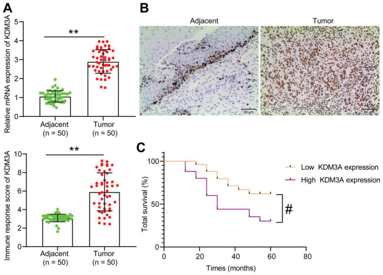
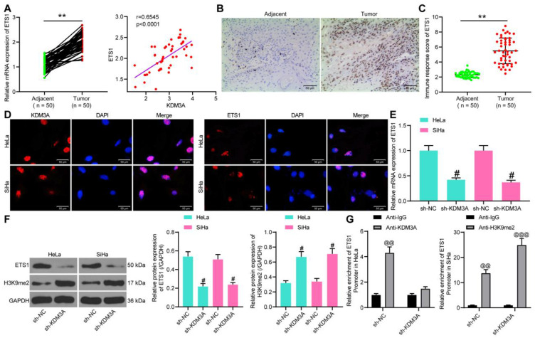
Lysine demethylase 3A () has been increasingly recognized as an important epigenetic regulator involved in cancer development. This study aims to explore the relevance of to cervical cancer (CC) progression and the molecules involved.
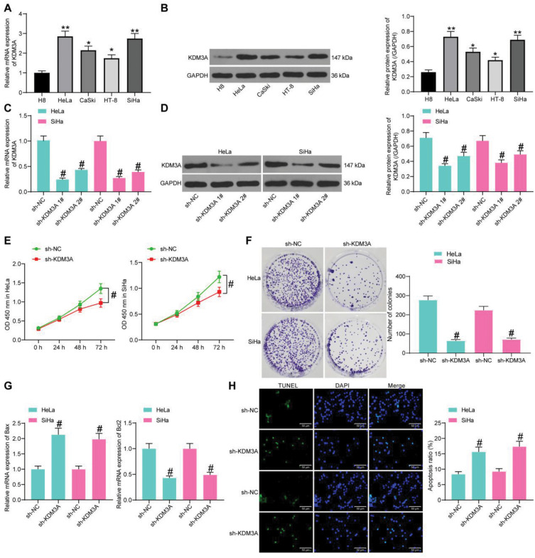
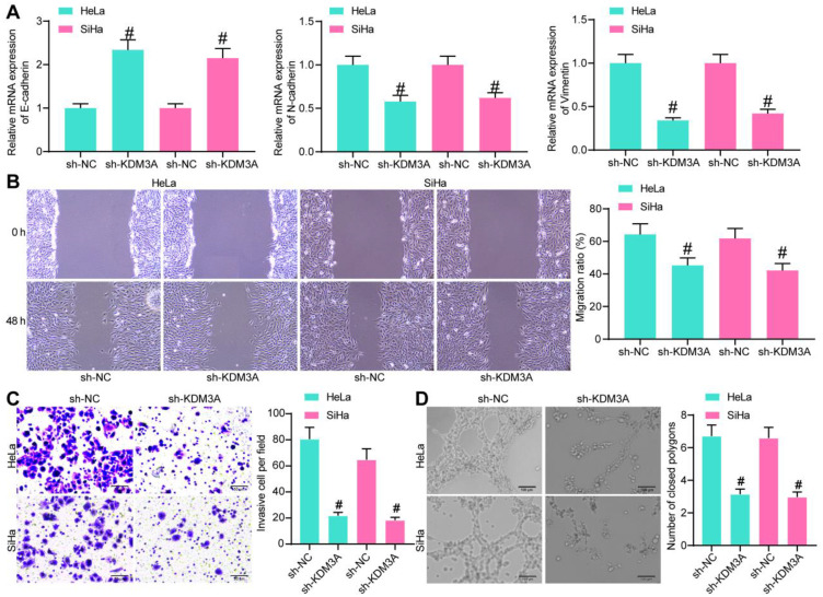
Tumor and the adjacent tissues from CC patients were collected. expression in tissues and CC cell lines and its correlation with the survival and prognosis of patients were determined. Malignant potentials of CC cells and the angiogenesis ability of HUVECs were measured to evaluate the function of on CC progression. The interactions among , H3K9me2 and , and the binding between and were validated through ChIP and luciferase assays. Altered expression of and was introduced to explore their roles in CC development.
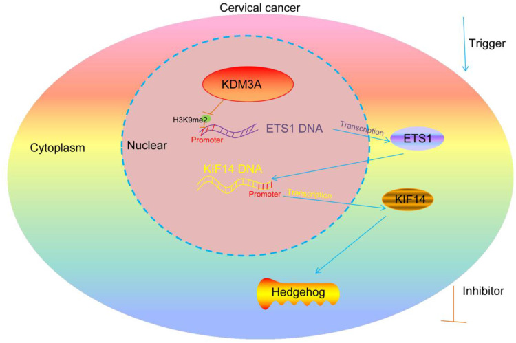
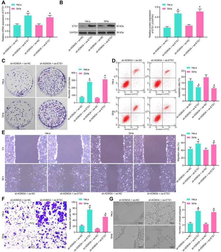
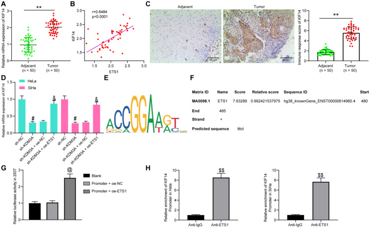
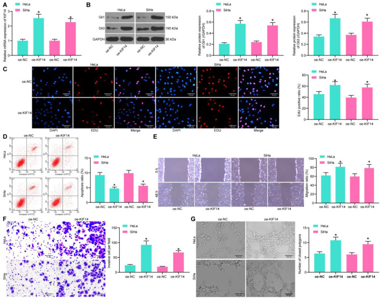
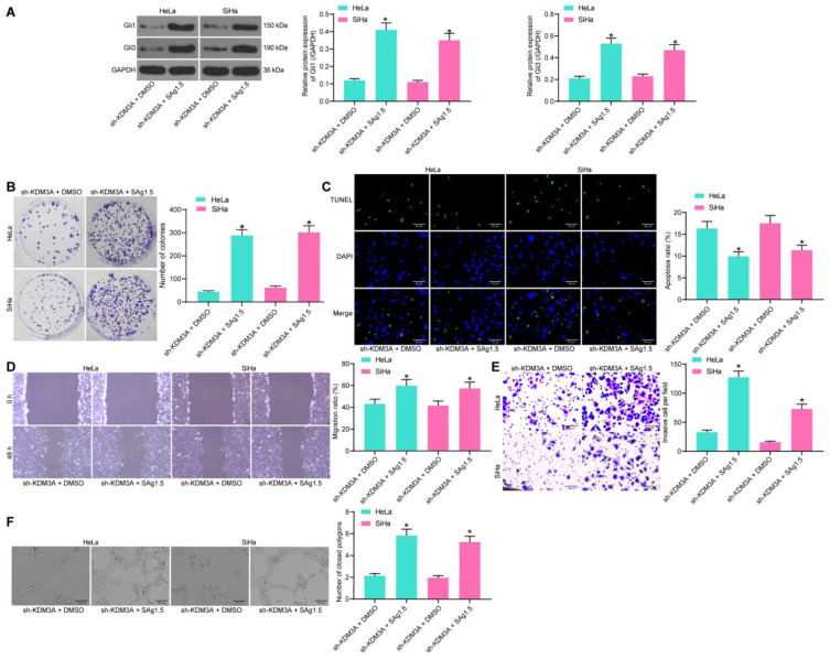
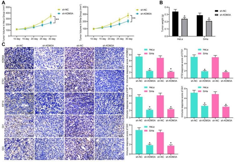
was abundantly expressed in CC tissues and cells and linked to dismal prognosis of CC patients. Knockdown of suppressed malignant behaviors of CC cells. was found to increase expression through the demethylation of H3K9me2. Overexpression of blocked the inhibiting roles of sh-KDM3A. could bind to the promoter region of to trigger its transcription. Overexpression ofaggravated the malignant behaviors of CC cells and the angiogenesis ability of HUVECs, and it activated the Hedgehog signaling pathway. Artificial activation of Hedgehog by Sag1.5 diminished the effects of sh-KDM3A. These changes were reproduced in vivo.
This study evidenced that promotes -mediated transcription to promote CC progression with the involvement of the Hedgehog activation.
赖氨酸去甲基化酶3A(KDM3A)已越来越被认为是参与癌症发展的重要表观遗传调节因子。本研究旨在探讨KDM3A与宫颈癌(CC)进展的相关性及相关分子。
收集CC患者的肿瘤组织及癌旁组织。检测KDM3A在组织和CC细胞系中的表达及其与患者生存和预后的相关性。检测CC细胞的恶性潜能及人脐静脉内皮细胞(HUVECs)的血管生成能力,以评估KDM3A对CC进展的作用。通过染色质免疫沉淀(ChIP)和荧光素酶测定验证KDM3A、H3K9me2和GLI1之间的相互作用以及KDM3A与GLI1启动子的结合。引入KDM3A和GLI1表达的改变以探讨它们在CC发展中的作用。
KDM3A在CC组织和细胞中高表达,与CC患者的不良预后相关。敲低KDM3A可抑制CC细胞的恶性行为。发现KDM3A通过使H3K9me2去甲基化增加GLI1表达。GLI1过表达可阻断sh-KDM3A的抑制作用。KDM3A可与GLI1的启动子区域结合以触发其转录。GLI1过表达加剧了CC细胞的恶性行为及HUVECs的血管生成能力,并激活了Hedgehog信号通路。Sag1.5人工激活Hedgehog可减弱sh-KDM3A的作用。这些变化在体内也得到了证实。
本研究证明KDM3A通过激活Hedgehog促进GLI1介导的转录,从而促进CC进展。