Yang Mingming, Dart Caroline, Kamishima Tomoko, Quayle John M
Department of Cardiology, Zhongda Hospital, Medical School of Southeast University, Nanjing, People's Republic of China.
Department of Cellular and Molecular Physiology, Institute of Translational Medicine, Liverpool, UK.
PeerJ. 2020 Nov 10;8:e10344. doi: 10.7717/peerj.10344. eCollection 2020.
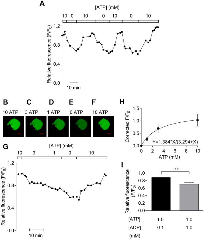
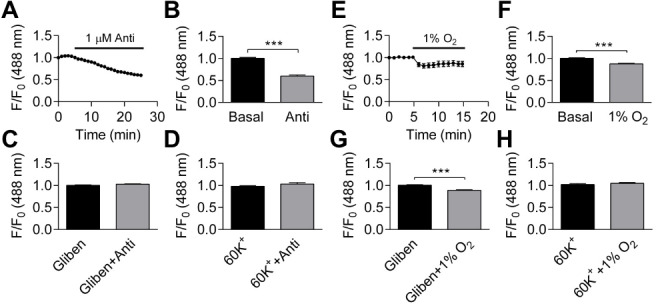
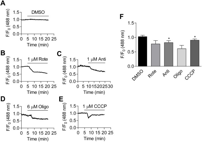
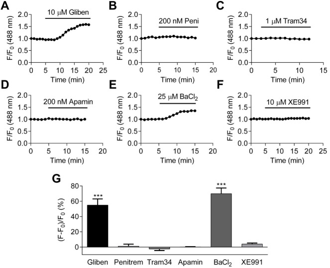
ATP-sensitive potassium (K) channels couple cellular metabolism to excitability, making them ideal candidate sensors for hypoxic vasodilation. However, it is still unknown whether cellular nucleotide levels are affected sufficiently to activate vascular K channels during hypoxia. To address this fundamental issue, we measured changes in the intracellular ATP:ADP ratio using the biosensors Perceval/PercevalHR, and membrane potential using the fluorescent probe DiBAC(3) in human coronary artery smooth muscle cells (HCASMCs). ATP:ADP ratio was significantly reduced by exposure to hypoxia. Application of metabolic inhibitors for oxidative phosphorylation also reduced ATP:ADP ratio. Hyperpolarization caused by inhibiting oxidative phosphorylation was blocked by either 10 µM glibenclamide or 60 mM K. Hyperpolarization caused by hypoxia was abolished by 60 mM K but not by individual K channel inhibitors. Taken together, these results suggest hypoxia causes hyperpolarization in part by modulating K channels in SMCs.
ATP 敏感性钾(K)通道将细胞代谢与兴奋性联系起来,使其成为缺氧性血管舒张的理想候选传感器。然而,细胞核苷酸水平在缺氧期间是否受到足够影响以激活血管 K 通道仍不清楚。为了解决这个基本问题,我们使用生物传感器 Perceval/PercevalHR 测量了人冠状动脉平滑肌细胞(HCASMCs)中细胞内 ATP:ADP 比值的变化,并使用荧光探针 DiBAC(3) 测量了膜电位。暴露于缺氧状态下,ATP:ADP 比值显著降低。应用氧化磷酸化代谢抑制剂也降低了 ATP:ADP 比值。抑制氧化磷酸化引起的超极化被 10 µM 格列本脲或 60 mM K 阻断。缺氧引起的超极化被 60 mM K 消除,但未被单个 K 通道抑制剂消除。综上所述,这些结果表明缺氧部分通过调节平滑肌细胞中的 K 通道导致超极化。