National Heart, Lung and Blood Institute (NHLBI), National Institutes of Health, 9000 Rockville Pike, Bethesda, MD, 20892, USA.
Cardiovascular Branch, NHLBI, 10 Center Drive, CRC, Room 5-5140, Bethesda, MD, 20892, USA.
J Nucl Cardiol. 2021 Oct;28(5):2033-2045. doi: 10.1007/s12350-020-02439-z. Epub 2020 Nov 26.
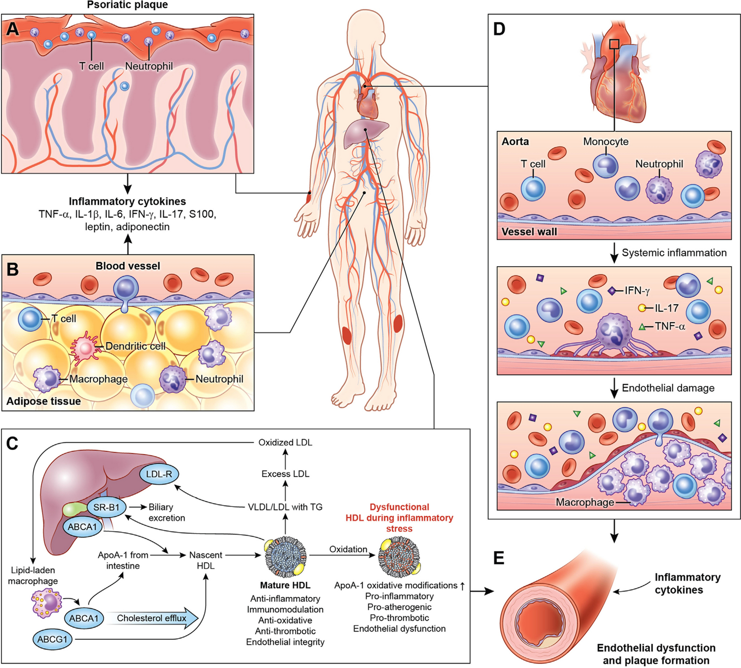
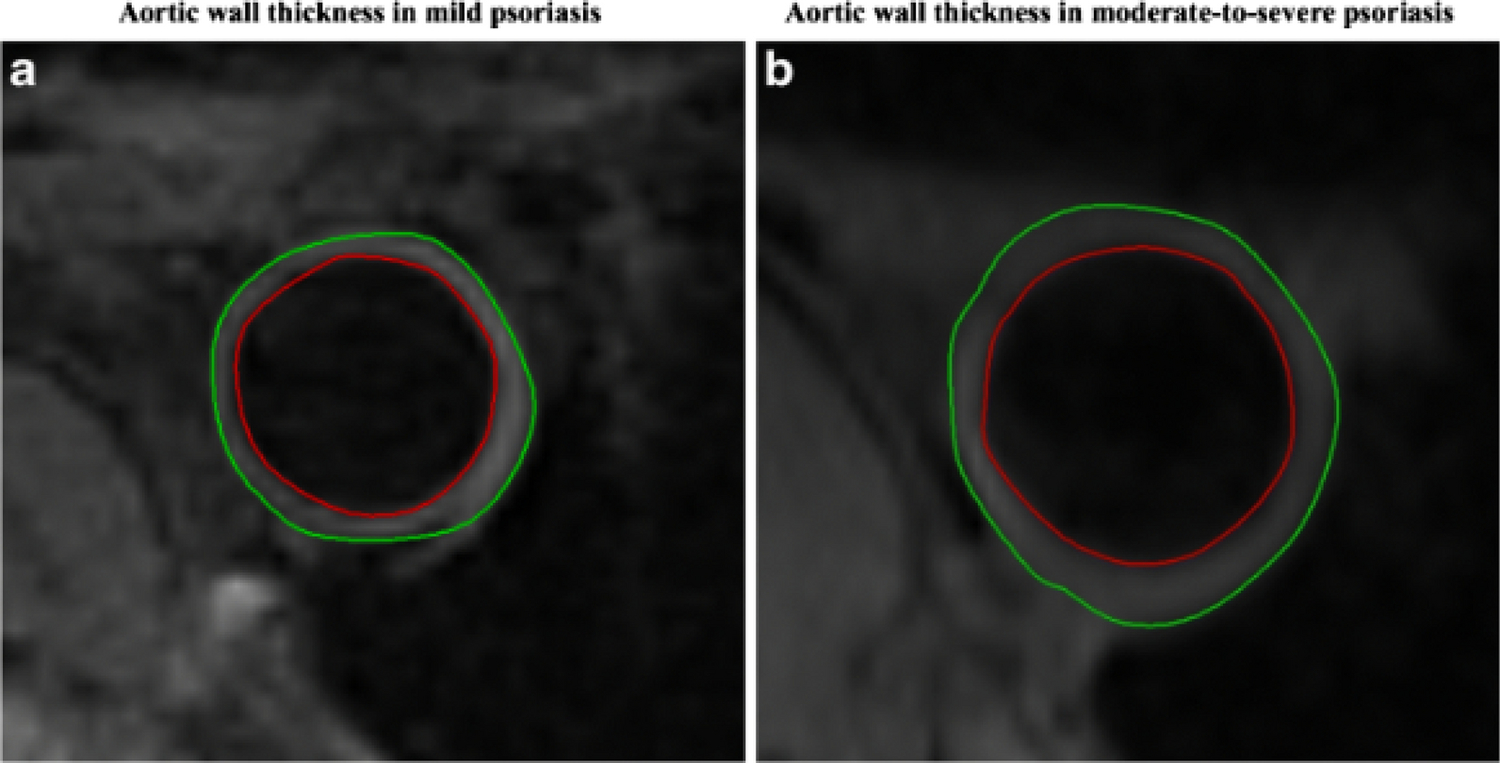
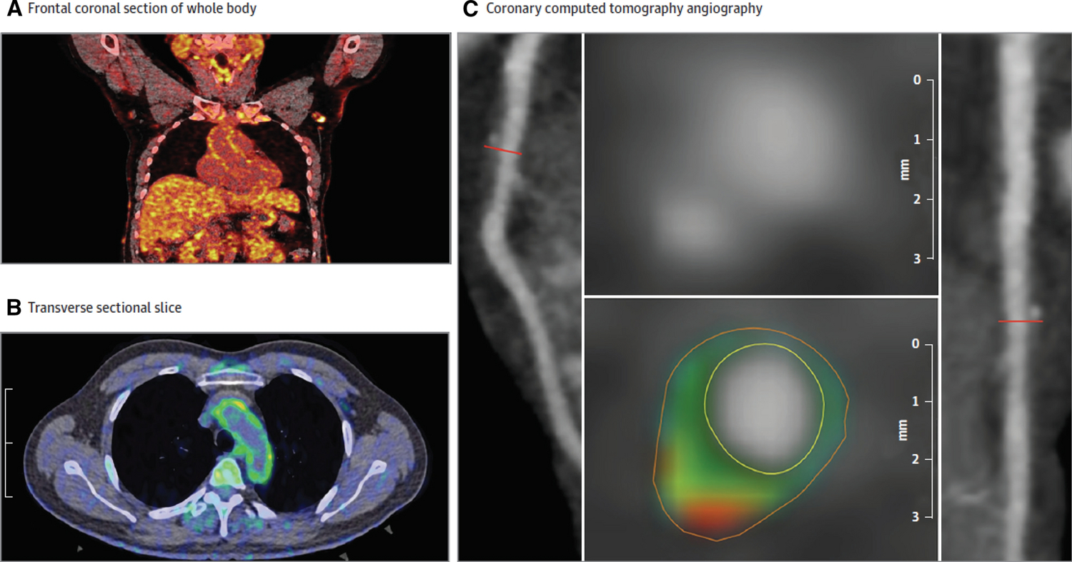
Over the past several decades, molecular imaging techniques to assess cellular processes in vivo have been integral in advancing our understanding of disease pathogenesis. F-fluorodeoxyglucose (18-FDG) positron emission tomography (PET) imaging in particular has shaped the field of atherosclerosis research by highlighting the importance of underlying inflammatory processes that are responsible for driving disease progression. The ability to assess physiology using molecular imaging, combining it with anatomic delineation using cardiac coronary angiography (CCTA) and magnetic resonance imaging (MRI) and lab-based techniques, provides a powerful combination to advance both research and ultimately clinical care. In this review, we demonstrate how molecular imaging studies, specifically using 18-FDG PET, have revealed that early vascular disease is a systemic process with multiple, concurrent biological mechanisms using inflammatory diseases as a basis to understand early atherosclerotic mechanisms in humans.
在过去的几十年中,评估体内细胞过程的分子成像技术在推进我们对疾病发病机制的理解方面发挥了重要作用。特别是 F-氟脱氧葡萄糖(18-FDG)正电子发射断层扫描(PET)成像通过强调负责驱动疾病进展的潜在炎症过程的重要性,塑造了动脉粥样硬化研究领域。使用分子成像评估生理学的能力,结合心脏冠状动脉造影(CCTA)和磁共振成像(MRI)以及基于实验室的技术进行解剖描绘,为推进研究并最终提供临床护理提供了强大的组合。在这篇综述中,我们展示了分子成像研究,特别是使用 18-FDG PET,如何揭示早期血管疾病是一种全身性疾病,具有多种并发生物学机制,使用炎症性疾病作为理解人类早期动脉粥样硬化机制的基础。