• 文献检索
  • 文档翻译
  • 深度研究
  • 学术资讯
  • Suppr Zotero 插件Zotero 插件
  • 邀请有礼
  • 套餐&价格
  • 历史记录
应用&插件
Suppr Zotero 插件Zotero 插件浏览器插件Mac 客户端Windows 客户端微信小程序
定价
高级版会员购买积分包购买API积分包
服务
文献检索文档翻译深度研究API 文档MCP 服务
关于我们
关于 Suppr公司介绍联系我们用户协议隐私条款
关注我们

Suppr 超能文献

核心技术专利:CN118964589B侵权必究
粤ICP备2023148730 号-1Suppr @ 2026

文献检索

告别复杂PubMed语法,用中文像聊天一样搜索,搜遍4000万医学文献。AI智能推荐,让科研检索更轻松。

立即免费搜索

文件翻译

保留排版,准确专业,支持PDF/Word/PPT等文件格式,支持 12+语言互译。

免费翻译文档

深度研究

AI帮你快速写综述,25分钟生成高质量综述,智能提取关键信息,辅助科研写作。

立即免费体验

利用节跃和混合技术的宽视场光谱成像及位移激发喇曼差光谱学。

Wide Field Spectral Imaging with Shifted Excitation Raman Difference Spectroscopy Using the Nod and Shuffle Technique.

机构信息

Leibniz Institute of Photonic Technology (Leibniz IPHT), Member of Research Alliance "Health Technologies", Albert-Einstein-Straße 9, 07743 Jena, Germany.

Leibniz Institute for Astrophysics Potsdam (AIP), Associated Member of Research Alliance "Health Technologies", An der Sternwarte 16, 14482 Potsdam, Germany.

出版信息

Sensors (Basel). 2020 Nov 24;20(23):6723. doi: 10.3390/s20236723.

DOI:10.3390/s20236723
PMID:33255459
原文链接:https://pmc.ncbi.nlm.nih.gov/articles/PMC7727830/
Abstract

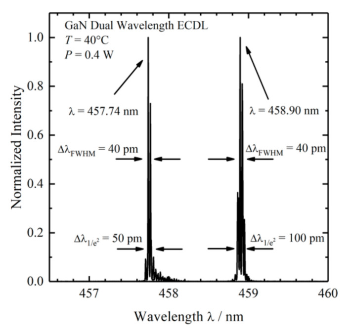
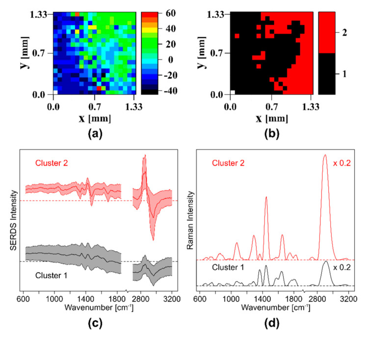
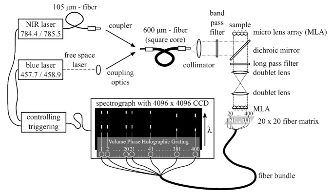
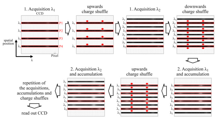
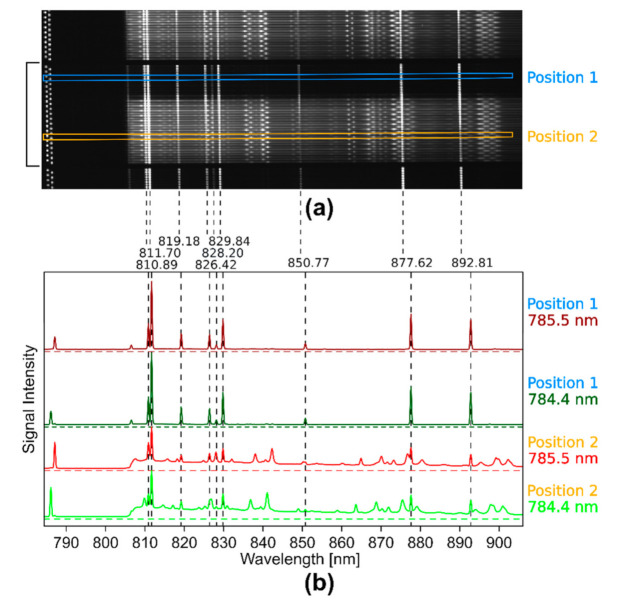
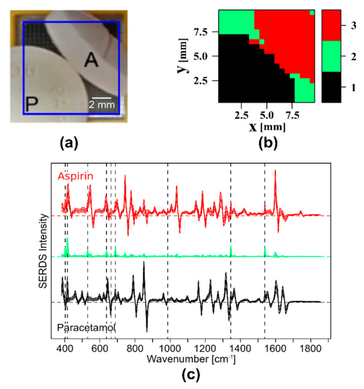
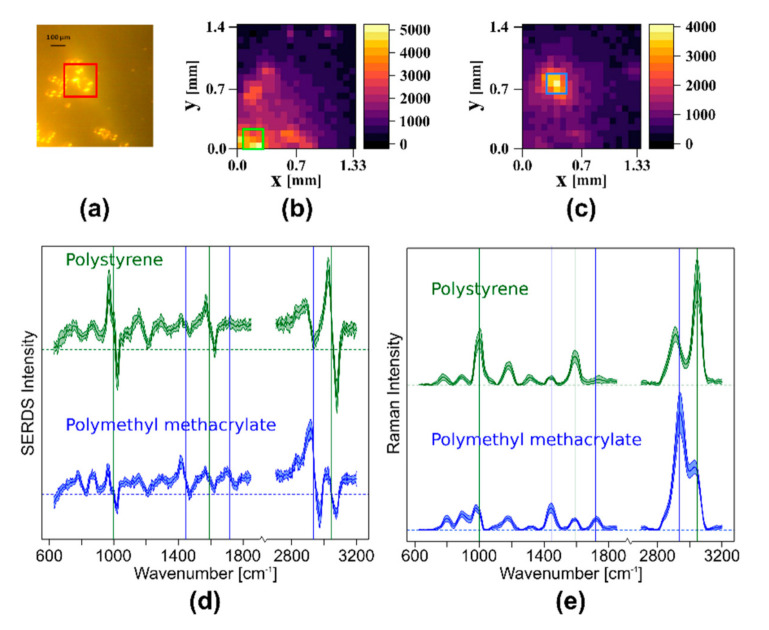
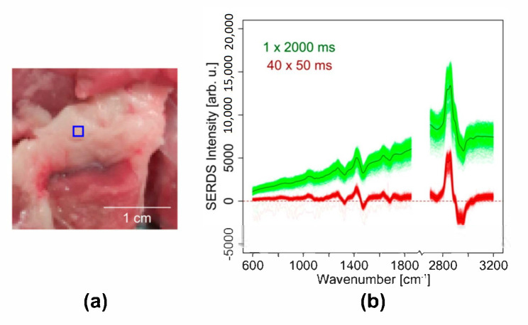
Wide field Raman imaging using the integral field spectroscopy approach was used as a fast, one shot imaging method for the simultaneous collection of all spectra composing a Raman image. For the suppression of autofluorescence and background signals such as room light, shifted excitation Raman difference spectroscopy (SERDS) was applied to remove background artifacts in Raman spectra. To reduce acquisition times in wide field SERDS imaging, we adapted the nod and shuffle technique from astrophysics and implemented it into a wide field SERDS imaging setup. In our adapted version, the nod corresponds to the change in excitation wavelength, whereas the shuffle corresponds to the shifting of charges up and down on a Charge-Coupled Device (CCD) chip synchronous to the change in excitation wavelength. We coupled this improved wide field SERDS imaging setup to diode lasers with 784.4/785.5 and 457.7/458.9 nm excitation and applied it to samples such as paracetamol and aspirin tablets, polystyrene and polymethyl methacrylate beads, as well as pork meat using multiple accumulations with acquisition times in the range of 50 to 200 ms. The results tackle two main challenges of SERDS imaging: gradual photobleaching changes the autofluorescence background, and multiple readouts of CCD detector prolong the acquisition time.

摘要

利用积分光谱学方法进行宽场 Raman 成像,作为一种快速的单次成像方法,可同时收集构成 Raman 图像的所有光谱。为了抑制自发荧光和背景信号(如室内光线),应用位移激发 Raman 差谱(SERDS)来去除 Raman 光谱中的背景伪影。为了减少宽场 SERDS 成像中的采集时间,我们从天体物理学中采用了“节”和“混洗”技术,并将其应用于宽场 SERDS 成像设置中。在我们的改编版本中,“节”对应于激发波长的变化,而“混洗”对应于电荷在 CCD 芯片上上下移动,与激发波长的变化同步。我们将这种改进的宽场 SERDS 成像设置与具有 784.4/785.5nm 和 457.7/458.9nm 激发的二极管激光器耦合,并将其应用于扑热息痛和阿司匹林药片、聚苯乙烯和聚甲基丙烯酸甲酯珠以及猪肉等样品,通过多次累积,采集时间在 50 到 200 毫秒之间。结果解决了 SERDS 成像的两个主要挑战:逐渐的光漂白会改变自发荧光背景,而 CCD 探测器的多次读数会延长采集时间。

https://cdn.ncbi.nlm.nih.gov/pmc/blobs/419c/7727830/26d7ef5310c7/sensors-20-06723-g008.jpg
https://cdn.ncbi.nlm.nih.gov/pmc/blobs/419c/7727830/e591f77e9713/sensors-20-06723-g001.jpg
https://cdn.ncbi.nlm.nih.gov/pmc/blobs/419c/7727830/870a47f08477/sensors-20-06723-g002.jpg
https://cdn.ncbi.nlm.nih.gov/pmc/blobs/419c/7727830/0df4edff7c89/sensors-20-06723-g003.jpg
https://cdn.ncbi.nlm.nih.gov/pmc/blobs/419c/7727830/5b5c5ba94b35/sensors-20-06723-g004.jpg
https://cdn.ncbi.nlm.nih.gov/pmc/blobs/419c/7727830/fe3510bb27cb/sensors-20-06723-g005.jpg
https://cdn.ncbi.nlm.nih.gov/pmc/blobs/419c/7727830/daa6ec957500/sensors-20-06723-g006.jpg
https://cdn.ncbi.nlm.nih.gov/pmc/blobs/419c/7727830/6a56f4203896/sensors-20-06723-g007.jpg
https://cdn.ncbi.nlm.nih.gov/pmc/blobs/419c/7727830/26d7ef5310c7/sensors-20-06723-g008.jpg

相似文献

1
Wide Field Spectral Imaging with Shifted Excitation Raman Difference Spectroscopy Using the Nod and Shuffle Technique.利用节跃和混合技术的宽视场光谱成像及位移激发喇曼差光谱学。
Sensors (Basel). 2020 Nov 24;20(23):6723. doi: 10.3390/s20236723.
2
Shifted Excitation Raman Difference Spectroscopy with Charge-Shifting Charge-Coupled Device (CCD) Lock-In Detection.移频激发拉曼差谱与电荷转移电荷耦合器件(CCD)锁相检测。
Appl Spectrosc. 2019 Nov;73(11):1265-1276. doi: 10.1177/0003702819859352. Epub 2019 Aug 12.
3
10 kHz Shifted-Excitation Raman Difference Spectroscopy with Charge-Shifting Charge-Coupled Device Read-Out for Effective Mitigation of Dynamic Interfering Backgrounds.10 kHz 频移激发拉曼差谱技术与电荷转移电荷耦合器件读出相结合,可有效减轻动态干扰背景。
Appl Spectrosc. 2023 Jun;77(6):569-582. doi: 10.1177/00037028231167441. Epub 2023 Apr 25.
4
Assessment of shifted excitation Raman difference spectroscopy in highly fluorescent biological samples.评估高荧光生物样本中的位移激发拉曼差光谱技术。
Analyst. 2021 Nov 8;146(22):6760-6767. doi: 10.1039/d1an01376a.
5
Parallelized shifted-excitation Raman difference spectroscopy for fluorescence rejection in a temporary varying system.用于临时变系统中荧光消除的并行移频共振拉曼差谱法。
J Biophotonics. 2019 Dec;12(12):e201960028. doi: 10.1002/jbio.201960028. Epub 2019 Aug 28.
6
Compact Handheld Probe for Shifted Excitation Raman Difference Spectroscopy with Implemented Dual-Wavelength Diode Laser at 785 Nanometers.紧凑型手持式探头,用于785纳米双波长二极管激光器实现的位移激发拉曼差分光谱分析。
Appl Spectrosc. 2015 Oct;69(10):1144-51. doi: 10.1366/15-07858.
7
Spectral reconstruction for shifted-excitation Raman difference spectroscopy (SERDS).频移激发拉曼差谱(SERDS)的光谱重建。
Talanta. 2018 Aug 15;186:372-380. doi: 10.1016/j.talanta.2018.04.050. Epub 2018 Apr 22.
8
Light-emitting diode based shifted-excitation Raman difference spectroscopy (LED-SERDS).基于发光二极管的位移激发拉曼差光谱(LED-SERDS)。
Analyst. 2013 Nov 7;138(21):6258-61. doi: 10.1039/c3an01367g.
9
Shifted Excitation Raman Difference Spectroscopy Combined with Wide Area Illumination and Sample Rotation for Wood Species Classification.位移激发拉曼差谱结合大面积照明和样品旋转用于木材种类分类。
Appl Spectrosc. 2023 Jun;77(6):666-681. doi: 10.1177/00037028231168405. Epub 2023 May 16.
10
Capability of shifted excitation Raman difference spectroscopy under ambient daylight.环境日光下位移激发拉曼差分光谱的能力
Appl Opt. 2015 Jun 10;54(17):5520-4. doi: 10.1364/AO.54.005520.

引用本文的文献

1
Raman difference spectroscopy and U-Net convolutional neural network for molecular analysis of cutaneous neurofibroma.拉曼差分光谱和 U-Net 卷积神经网络在皮肤神经纤维瘤的分子分析中的应用。
PLoS One. 2024 Apr 11;19(4):e0302017. doi: 10.1371/journal.pone.0302017. eCollection 2024.
2
Optics miniaturization strategy for demanding Raman spectroscopy applications.适用于高要求拉曼光谱应用的光学小型化策略。
Nat Commun. 2024 Apr 8;15(1):3049. doi: 10.1038/s41467-024-47044-7.
3
Compact Shortwave Infrared Imaging Spectrometer Based on a Catadioptric Prism.

本文引用的文献

1
New methodology to process shifted excitation Raman difference spectroscopy data: a case study of pollen classification.新方法处理偏移激发拉曼差光谱数据:花粉分类的案例研究。
Sci Rep. 2020 Jul 8;10(1):11215. doi: 10.1038/s41598-020-67897-4.
2
Molecular origin of the Raman signal from Aspergillus nidulans conidia and observation of fluorescence vibrational structure at room temperature.从aspergillus nidulans 分生孢子中获得的 Raman 信号的分子起源及室温下荧光振动结构的观察。
Sci Rep. 2020 Mar 25;10(1):5428. doi: 10.1038/s41598-020-62112-w.
3
Bladder tissue characterization using probe-based Raman spectroscopy: Evaluation of tissue heterogeneity and influence on the model prediction.
基于折反射棱镜的紧凑型短波红外成像光谱仪。
Sensors (Basel). 2022 Jun 18;22(12):4611. doi: 10.3390/s22124611.
4
Looking for a perfect match: multimodal combinations of Raman spectroscopy for biomedical applications.寻找完美匹配:用于生物医学应用的拉曼光谱学的多模式组合。
J Biomed Opt. 2021 Aug;26(8). doi: 10.1117/1.JBO.26.8.080601.
基于探针的拉曼光谱法对膀胱组织的表征:组织异质性评估及其对模型预测的影响。
J Biophotonics. 2020 Feb;13(2):e201960025. doi: 10.1002/jbio.201960025. Epub 2019 Dec 2.
4
Raman spectroscopy for rapid intra-operative margin analysis of surgically excised tumour specimens.拉曼光谱法快速分析手术切除的肿瘤标本的术中边缘。
Analyst. 2019 Nov 4;144(22):6479-6496. doi: 10.1039/c9an01163c.
5
Shifted Excitation Raman Difference Spectroscopy with Charge-Shifting Charge-Coupled Device (CCD) Lock-In Detection.移频激发拉曼差谱与电荷转移电荷耦合器件(CCD)锁相检测。
Appl Spectrosc. 2019 Nov;73(11):1265-1276. doi: 10.1177/0003702819859352. Epub 2019 Aug 12.
6
Applications of Spatial Light Modulators in Raman Spectroscopy.空间光调制器在拉曼光谱学中的应用。
Appl Spectrosc. 2019 Jul;73(7):727-746. doi: 10.1177/0003702819834575. Epub 2019 Apr 16.
7
Nonscanning large-area Raman imaging for ex vivo/in vivo skin cancer discrimination.非扫描大面积拉曼成像用于离体/体内皮肤癌鉴别。
J Biomed Opt. 2018 Oct;23(10):1-11. doi: 10.1117/1.JBO.23.10.105001.
8
Breast Tumor Analysis Using Shifted-Excitation Raman Difference Spectroscopy (SERDS).使用位移激发拉曼差分光谱(SERDS)进行乳腺肿瘤分析。
Technol Cancer Res Treat. 2018 Jan 1;17:1533033818782532. doi: 10.1177/1533033818782532.
9
A new method using Raman spectroscopy for in vivo targeted brain cancer tissue biopsy.一种使用拉曼光谱进行体内靶向脑癌组织活检的新方法。
Sci Rep. 2018 Jan 29;8(1):1792. doi: 10.1038/s41598-018-20233-3.
10
Evaluation of Shifted Excitation Raman Difference Spectroscopy and Comparison to Computational Background Correction Methods Applied to Biochemical Raman Spectra.移位激发拉曼差谱法的评价及与应用于生化拉曼光谱的计算背景校正方法的比较。
Sensors (Basel). 2017 Jul 27;17(8):1724. doi: 10.3390/s17081724.