Fernandez Sandra V, MacFarlane Alexander W, Jillab Mowafaq, Arisi Maria F, Yearley Jennifer, Annamalai Lakshmanan, Gong Yulan, Cai Kathy Q, Alpaugh R Katherine, Cristofanilli Massimo, Campbell Kerry S
Department of Medical Oncology, Fox Chase Cancer Center, Philadelphia, PA, 19111, USA.
Blood Cell Development and Function Program, Institute for Cancer Research, Fox Chase Cancer Center, 333 Cottman Ave, Philadelphia, PA, 19111, USA.
Breast Cancer Res. 2020 Dec 2;22(1):134. doi: 10.1186/s13058-020-01371-x.
Inflammatory breast cancer (IBC) is a rare but aggressive carcinoma characterized by severe erythema and edema of the breast, with many patients presenting in advanced metastatic disease. The "inflammatory" nature is not due to classic immune-mediated inflammation, but instead results from tumor-mediated blockage of dermal lymphatic ducts. Previous work has shown that expression of PD-L1 on tumor cells can suppress T cell activation in triple-negative (TN) non-IBC breast cancer. In the present work, we investigated immune parameters in peripheral blood of metastatic IBC patients to determine whether cellular components of the immune system are altered, thereby contributing to pathogenesis of the disease. These immune parameters were also compared to PD-1 and PD-L1 expression in IBC tumor biopsies.
Flow cytometry-based immune phenotyping was performed using fresh peripheral blood from 14 stage IV IBC patients and compared to 11 healthy age-similar control women. Immunohistochemistry for CD20, CD3, PD-1, and PD-L1 was performed on tumor biopsies of these metastatic IBC patients.
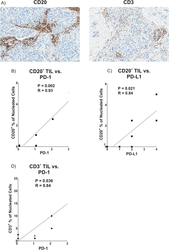
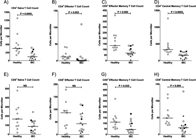
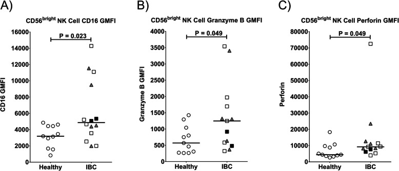
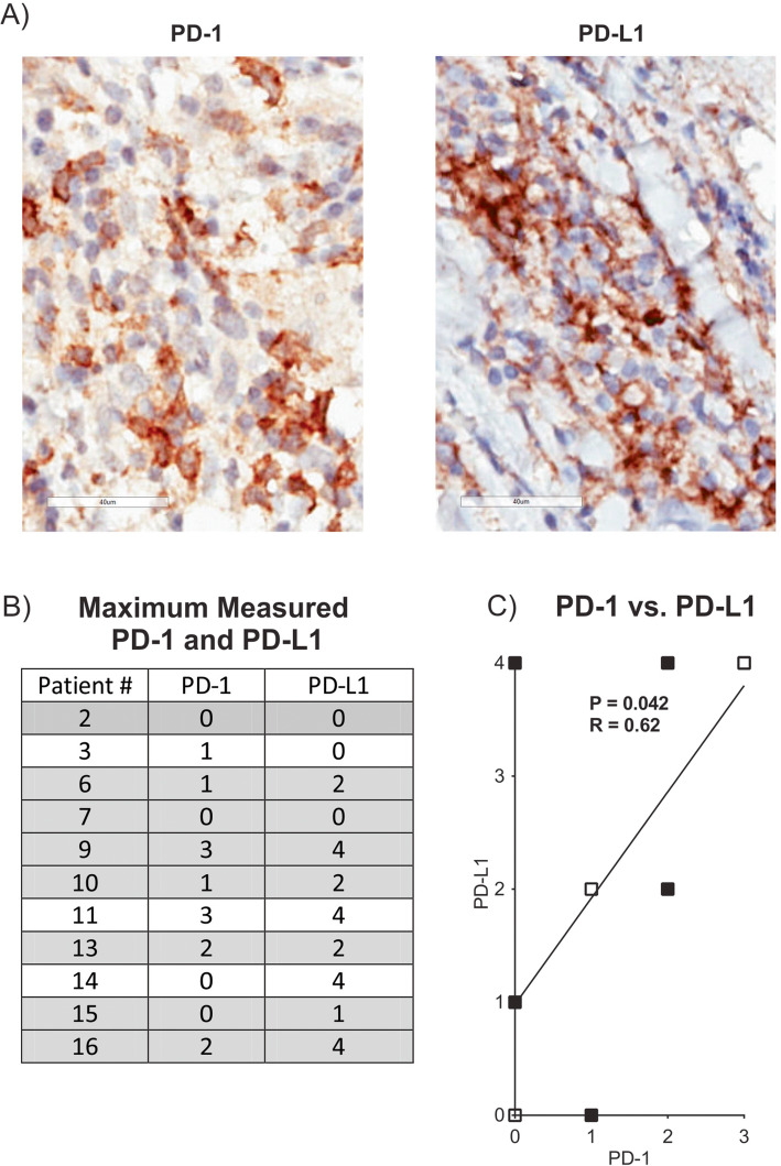
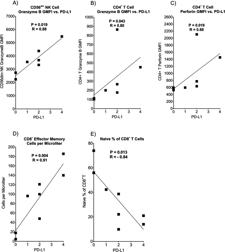
IBC patients with Stage IV disease had lymphopenia with significant reductions in circulating T, B, and NK cells. Reductions were observed in all subsets of CD4 T cells, whereas reductions in CD8 T cells were more concentrated in memory subsets. Immature cytokine-producing CD56 NK cells expressed higher levels of FcγRIIIa and cytolytic granule components, suggesting accelerated maturation to cytolytic CD56 cells. Immunohistochemical analysis of tumor biopsies demonstrated moderate to high expression of PD-1 in 18.2% of patients and of PD-L1 in 36.4% of patients. Interestingly, a positive correlation was observed between co-expression levels of PD-L1 and PD-1 in tumor biopsies, and higher expression of PD-L1 in tumor biopsies correlated with higher expression of cytolytic granule components in blood CD4 T cells and CD56 NK cells, and higher numbers of CD8 effector memory T cells in peripheral blood. PD-1 expression in tumor also correlated with increased infiltration of CD20 B cells in the tumor.
Our results suggest that while lymphocyte populations are severely compromised in stage IV IBC patients, an immune response toward the tumor had occurred in some patients, providing biological rationale to evaluate PD-1/PD-L1 immunotherapies for IBC.
炎性乳腺癌(IBC)是一种罕见但侵袭性强的癌症,其特征为乳房严重红斑和水肿,许多患者就诊时已处于晚期转移性疾病阶段。其“炎性”本质并非源于经典的免疫介导炎症,而是肿瘤介导的真皮淋巴管阻塞所致。先前的研究表明,肿瘤细胞上PD-L1的表达可抑制三阴性(TN)非IBC乳腺癌中的T细胞活化。在本研究中,我们调查了转移性IBC患者外周血中的免疫参数,以确定免疫系统的细胞成分是否发生改变,从而有助于该疾病的发病机制。这些免疫参数还与IBC肿瘤活检中的PD-1和PD-L1表达进行了比较。
使用14例IV期IBC患者的新鲜外周血进行基于流式细胞术的免疫表型分析,并与11名年龄相仿的健康对照女性进行比较。对这些转移性IBC患者的肿瘤活检进行CD20、CD3、PD-1和PD-L1的免疫组织化学检测。
IV期疾病的IBC患者出现淋巴细胞减少,循环中的T、B和NK细胞显著减少。在所有CD4 T细胞亚群中均观察到减少,而CD8 T细胞的减少更集中在记忆亚群中。产生细胞因子的未成熟CD56 NK细胞表达较高水平的FcγRIIIa和溶细胞颗粒成分,表明加速成熟为溶细胞性CD56细胞。肿瘤活检的免疫组织化学分析显示,18.2%的患者PD-1呈中度至高度表达,36.4%的患者PD-L1呈中度至高度表达。有趣的是,在肿瘤活检中观察到PD-L1和PD-1的共表达水平之间呈正相关,肿瘤活检中PD-L1的较高表达与血液CD4 T细胞和CD56 NK细胞中溶细胞颗粒成分的较高表达以及外周血中CD8效应记忆T细胞的数量增加相关。肿瘤中的PD-1表达也与肿瘤中CD20 B细胞浸润增加相关。
我们的结果表明,虽然IV期IBC患者的淋巴细胞群体严重受损,但部分患者已对肿瘤产生免疫反应,这为评估IBC的PD-1/PD-L1免疫疗法提供了生物学依据。