• 文献检索
  • 文档翻译
  • 深度研究
  • 学术资讯
  • Suppr Zotero 插件Zotero 插件
  • 邀请有礼
  • 套餐&价格
  • 历史记录
应用&插件
Suppr Zotero 插件Zotero 插件浏览器插件Mac 客户端Windows 客户端微信小程序
定价
高级版会员购买积分包购买API积分包
服务
文献检索文档翻译深度研究API 文档MCP 服务
关于我们
关于 Suppr公司介绍联系我们用户协议隐私条款
关注我们

Suppr 超能文献

核心技术专利:CN118964589B侵权必究
粤ICP备2023148730 号-1Suppr @ 2026

文献检索

告别复杂PubMed语法,用中文像聊天一样搜索,搜遍4000万医学文献。AI智能推荐,让科研检索更轻松。

立即免费搜索

文件翻译

保留排版,准确专业,支持PDF/Word/PPT等文件格式,支持 12+语言互译。

免费翻译文档

深度研究

AI帮你快速写综述,25分钟生成高质量综述,智能提取关键信息,辅助科研写作。

立即免费体验

对区域性脑容量测量的单个体分析可能会受到头部大小调整方法的强烈影响。

Single-subject analysis of regional brain volumetric measures can be strongly influenced by the method for head size adjustment.

机构信息

Jung Diagnostics GmbH, Hamburg, Germany.

Institute of Diagnostic and Interventional Neuroradiology, University Hospital Carl Gustav Carus, Technische Universität Dresden, Dresden, Germany.

出版信息

Neuroradiology. 2022 Oct;64(10):2001-2009. doi: 10.1007/s00234-022-02961-6. Epub 2022 Apr 25.

DOI:10.1007/s00234-022-02961-6
PMID:35462574
原文链接:https://pmc.ncbi.nlm.nih.gov/articles/PMC9474386/
Abstract

PURPOSE

Total intracranial volume (TIV) is often a nuisance covariate in MRI-based brain volumetry. This study compared two TIV adjustment methods with respect to their impact on z-scores in single subject analyses of regional brain volume estimates.

METHODS

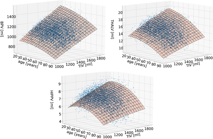
Brain parenchyma, hippocampus, thalamus, and TIV were segmented in a normal database comprising 5059 T1w images. Regional volume estimates were adjusted for TIV using the residual method or the proportion method. Age was taken into account by regression with both methods. TIV- and age-adjusted regional volumes were transformed to z-scores and then compared between the two adjustment methods. Their impact on the detection of thalamus atrophy was tested in 127 patients with multiple sclerosis.

RESULTS

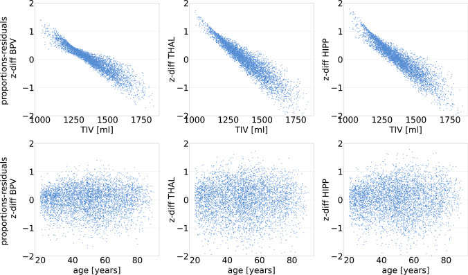
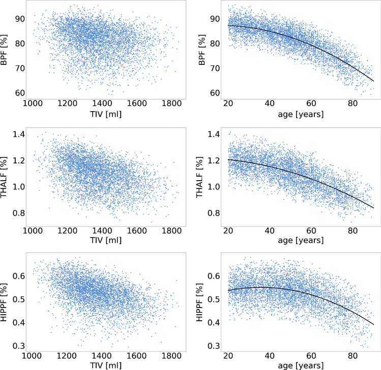
The residual method removed the association with TIV in all regions. The proportion method resulted in a switch of the direction without relevant change of the strength of the association. The reduction of physiological between-subject variability was larger with the residual method than with the proportion method. The difference between z-scores obtained with the residual method versus the proportion method was strongly correlated with TIV. It was larger than one z-score point in 5% of the subjects. The area under the ROC curve of the TIV- and age-adjusted thalamus volume for identification of multiple sclerosis patients was larger with the residual method than with the proportion method (0.84 versus 0.79).

CONCLUSION

The residual method should be preferred for TIV and age adjustments of T1w-MRI-based brain volume estimates in single subject analyses.

摘要

目的

全脑容量(TIV)通常是 MRI 脑容积测量中的一个干扰协变量。本研究比较了两种 TIV 调整方法对个体脑区容积估计的 z 分数的影响。

方法

在一个由 5059 个 T1w 图像组成的正常数据库中,对脑实质、海马体、丘脑和 TIV 进行分割。使用残差法或比例法对 TIV 进行调整,对 TIV 和年龄进行回归。将 TIV 和年龄调整后的区域容积转换为 z 分数,然后比较两种调整方法。在 127 名多发性硬化症患者中,测试了它们对丘脑萎缩检测的影响。

结果

残差法消除了所有区域与 TIV 的相关性。比例法导致了相关性的方向改变,而没有改变相关性的强度。残差法比比例法更能减少生理上的个体间变异性。与比例法相比,残差法获得的 z 分数之间的差异与 TIV 密切相关。在 5%的受试者中,这种差异大于一个 z 分数点。TIV 和年龄调整后的丘脑容积的 ROC 曲线下面积,残差法比比例法更大(0.84 比 0.79)。

结论

在个体脑区容积估计的 T1w-MRI 分析中,应优先使用残差法进行 TIV 和年龄调整。

https://cdn.ncbi.nlm.nih.gov/pmc/blobs/3efb/9474386/7e89dbe6fcdb/234_2022_2961_Fig3_HTML.jpg
https://cdn.ncbi.nlm.nih.gov/pmc/blobs/3efb/9474386/918cf37326e7/234_2022_2961_Fig1_HTML.jpg
https://cdn.ncbi.nlm.nih.gov/pmc/blobs/3efb/9474386/583c90532005/234_2022_2961_Fig2_HTML.jpg
https://cdn.ncbi.nlm.nih.gov/pmc/blobs/3efb/9474386/7e89dbe6fcdb/234_2022_2961_Fig3_HTML.jpg
https://cdn.ncbi.nlm.nih.gov/pmc/blobs/3efb/9474386/918cf37326e7/234_2022_2961_Fig1_HTML.jpg
https://cdn.ncbi.nlm.nih.gov/pmc/blobs/3efb/9474386/583c90532005/234_2022_2961_Fig2_HTML.jpg
https://cdn.ncbi.nlm.nih.gov/pmc/blobs/3efb/9474386/7e89dbe6fcdb/234_2022_2961_Fig3_HTML.jpg

相似文献

1
Single-subject analysis of regional brain volumetric measures can be strongly influenced by the method for head size adjustment.对区域性脑容量测量的单个体分析可能会受到头部大小调整方法的强烈影响。
Neuroradiology. 2022 Oct;64(10):2001-2009. doi: 10.1007/s00234-022-02961-6. Epub 2022 Apr 25.
2
Evaluating the Impact of Intracranial Volume Correction Approaches on the Quantification of Intracranial Structures in MRI: A Systematic Analysis.评估颅内容积校正方法对 MRI 中颅内结构定量分析的影响:系统分析。
J Magn Reson Imaging. 2024 Jun;59(6):2164-2177. doi: 10.1002/jmri.28974. Epub 2023 Sep 13.
3
A unified approach for morphometric and functional data analysis in young, old, and demented adults using automated atlas-based head size normalization: reliability and validation against manual measurement of total intracranial volume.一种使用基于自动图谱的头部大小归一化方法对年轻、老年和痴呆成年人的形态学和功能数据分析的统一方法:与手动测量总颅内体积相比的可靠性和验证。
Neuroimage. 2004 Oct;23(2):724-38. doi: 10.1016/j.neuroimage.2004.06.018.
4
Normalization of cerebral volumes by use of intracranial volume: implications for longitudinal quantitative MR imaging.使用颅内体积对脑容量进行标准化:对纵向定量磁共振成像的意义。
AJNR Am J Neuroradiol. 2001 Sep;22(8):1483-9.
5
Accurate automatic estimation of total intracranial volume: a nuisance variable with less nuisance.准确自动估计总颅内体积:一个麻烦程度较低的干扰变量。
Neuroimage. 2015 Jan 1;104:366-72. doi: 10.1016/j.neuroimage.2014.09.034. Epub 2014 Oct 1.
6
Choroid plexus volume in multiple sclerosis predicts expansion of chronic lesions and brain atrophy.脉络丛体积在多发性硬化症中可预测慢性病变和脑萎缩的扩展。
Ann Clin Transl Neurol. 2022 Oct;9(10):1528-1537. doi: 10.1002/acn3.51644. Epub 2022 Sep 3.
7
Correction for intracranial volume in analysis of whole brain atrophy in multiple sclerosis: the proportion vs. residual method.多发性硬化症全脑萎缩分析中颅内体积的校正:比例法与残差法
Neuroimage. 2004 Aug;22(4):1732-43. doi: 10.1016/j.neuroimage.2004.03.037.
8
Intracranial volume and Alzheimer disease: evidence against the cerebral reserve hypothesis.颅内体积与阿尔茨海默病:反对大脑储备假说的证据
Arch Neurol. 2000 Feb;57(2):220-4. doi: 10.1001/archneur.57.2.220.
9
Comparative analysis of methods of volume adjustment in hippocampal volumetry for the diagnosis of Alzheimer disease.比较分析海马体积测量中容积调整方法在阿尔茨海默病诊断中的应用。
J Neuroradiol. 2020 Mar;47(2):161-165. doi: 10.1016/j.neurad.2019.02.004. Epub 2019 Mar 8.
10
Sex differences in gray matter volume: how many and how large are they really?性别差异在大脑灰质体积上的表现:究竟有多少,又有多大?
Biol Sex Differ. 2019 Jul 1;10(1):32. doi: 10.1186/s13293-019-0245-7.

引用本文的文献

1
Lower hippocampal volumes at baseline are associated with higher volume loss in healthy elderly.基线时海马体体积较小与健康老年人更大的体积损失相关。
Front Aging Neurosci. 2025 Jul 16;17:1542857. doi: 10.3389/fnagi.2025.1542857. eCollection 2025.
2
Comparison of approaches to control for intracranial volume in research on the association of brain volumes with cognitive outcomes.脑容量与认知结果关联研究中控制颅内体积方法的比较。
Hum Brain Mapp. 2024 Mar;45(4):e26633. doi: 10.1002/hbm.26633.
3
Distinct cerebral small vessel disease impairment in early- and late-onset Alzheimer's disease.

本文引用的文献

1
Infratentorial lesions in multiple sclerosis patients: intra- and inter-rater variability in comparison to a fully automated segmentation using 3D convolutional neural networks.多发性硬化症患者的桥脑下病变:与使用 3D 卷积神经网络的全自动分割相比的组内和组间可变性。
Eur Radiol. 2022 Apr;32(4):2798-2809. doi: 10.1007/s00330-021-08329-3. Epub 2021 Oct 13.
2
Neuroanatomical norms in the UK Biobank: The impact of allometric scaling, sex, and age.英国生物银行中的神经解剖学规范: 比例缩放、性别和年龄的影响。
Hum Brain Mapp. 2021 Oct 1;42(14):4623-4642. doi: 10.1002/hbm.25572. Epub 2021 Jul 16.
3
Machine learning classifier to identify clinical and radiological features relevant to disability progression in multiple sclerosis.
早发性和晚发性阿尔茨海默病中不同的脑小血管病损害。
Ann Clin Transl Neurol. 2023 Aug;10(8):1326-1337. doi: 10.1002/acn3.51824. Epub 2023 Jun 22.
4
Commercial volumetric MRI reporting tools in multiple sclerosis: a systematic review of the evidence.商业容积磁共振成像报告工具在多发性硬化症中的应用:系统评价证据。
Neuroradiology. 2023 Jan;65(1):5-24. doi: 10.1007/s00234-022-03074-w. Epub 2022 Nov 4.
机器学习分类器,用于识别多发性硬化症中与残疾进展相关的临床和影像学特征。
J Neurol. 2021 Dec;268(12):4834-4845. doi: 10.1007/s00415-021-10605-7. Epub 2021 May 10.
4
Thalamic Injury and Cognition in Multiple Sclerosis.多发性硬化症中的丘脑损伤与认知
Front Neurol. 2021 Feb 5;11:623914. doi: 10.3389/fneur.2020.623914. eCollection 2020.
5
Age-dependent cut-offs for pathological deep gray matter and thalamic volume loss using Jacobian integration.基于雅可比积分的病理性深部灰质和丘脑体积损失的年龄依赖性截断值。
Neuroimage Clin. 2020;28:102478. doi: 10.1016/j.nicl.2020.102478. Epub 2020 Oct 27.
6
Fully automated longitudinal segmentation of new or enlarged multiple sclerosis lesions using 3D convolutional neural networks.使用 3D 卷积神经网络全自动纵向分割新的或扩大的多发性硬化病变。
Neuroimage Clin. 2020;28:102445. doi: 10.1016/j.nicl.2020.102445. Epub 2020 Sep 24.
7
Effects of different intracranial volume correction methods on univariate sex differences in grey matter volume and multivariate sex prediction.不同颅内容量校正方法对灰质体积单变量性别差异和多变量性别预测的影响。
Sci Rep. 2020 Jul 31;10(1):12953. doi: 10.1038/s41598-020-69361-9.
8
Whole Brain and Cranial Size Adjustments in Volumetric Brain Analyses of Sex- and Age-Related Trends.性别和年龄相关趋势的脑容量分析中的全脑和颅骨大小调整
Front Neurosci. 2020 Apr 3;14:278. doi: 10.3389/fnins.2020.00278. eCollection 2020.
9
Hippocampal Sclerosis Detection with NeuroQuant Compared with Neuroradiologists.神经定量分析与神经放射科医师检测海马硬化的比较。
AJNR Am J Neuroradiol. 2020 Apr;41(4):591-597. doi: 10.3174/ajnr.A6454. Epub 2020 Mar 26.
10
Sex differences in gray matter volume: how many and how large are they really?性别差异在大脑灰质体积上的表现:究竟有多少,又有多大?
Biol Sex Differ. 2019 Jul 1;10(1):32. doi: 10.1186/s13293-019-0245-7.