Irodi Aparna, Chacko Binita R, Prajapati Anand, Prabhu Anne J, Vimala Leena R, Christopher Devasahayam J, Gnanamuthu Birla R
Department of Radiology, Christian Medical College, Vellore, Tamil Nadu, India.
Department of Medical Imaging, Sunnybrook Health Sciences Centre, University of Toronto, M4N 3M5, Canada.
Indian J Radiol Imaging. 2020 Jul-Sep;30(3):266-272. doi: 10.4103/ijri.IJRI_93_20. Epub 2020 Oct 15.
Inflammatory myofibroblastic tumour (IMT) is a rare mesenchymal neoplasm with intermediate malignant potential. The aim of this study is to describe and compare the clinical presentation, computed tomography (CT) findings and anaplastic lymphoma kinase -1 (ALK-1) expression of IMT of the thorax in children and adults. We also sought to study the tumour behaviour after treatment on the follow-up imaging.
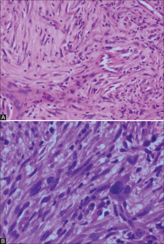
This is a retrospective observational study of 22 histopathologically proven cases of IMT in the thorax. The clinical parameters, CT findings, biopsy results, treatment received and follow-up were recorded. Statistical analysis was performed using Fisher's exact test.
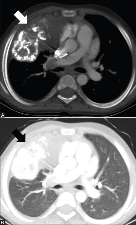
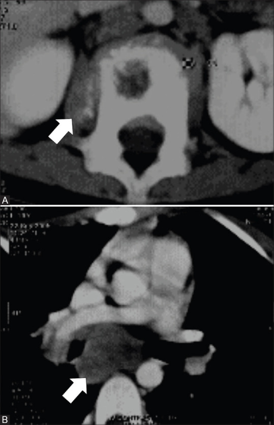
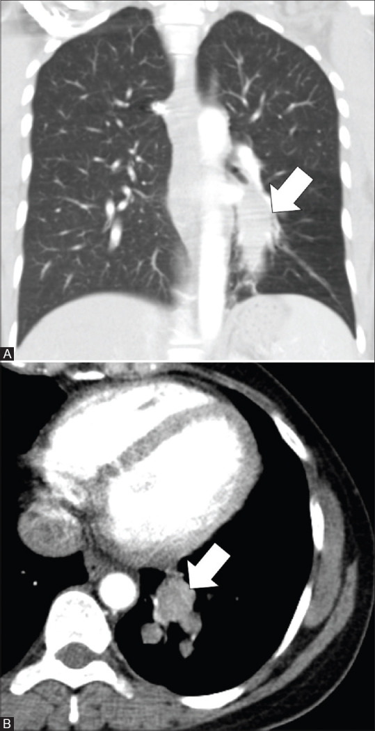
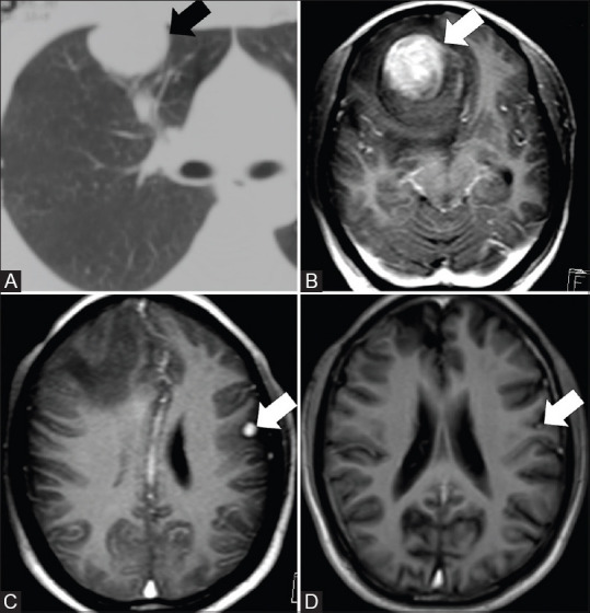
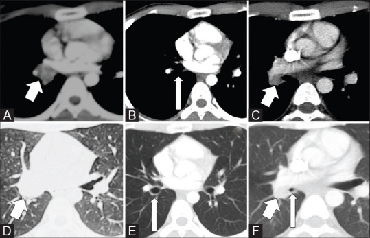
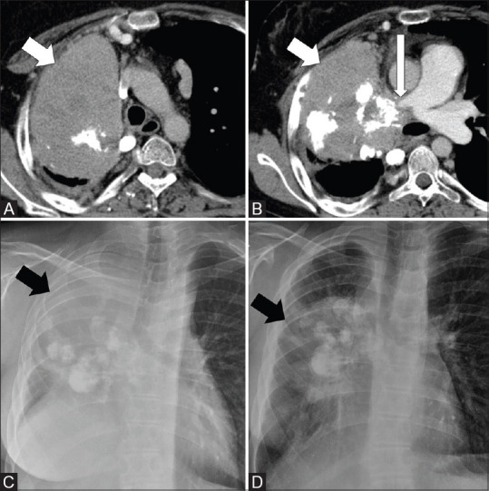
IMT of the thorax had diverse imaging appearances, presenting either as large invasive lung masses with or without calcifications or as smaller endobronchial lesions. Children commonly presented with long duration fever ( = 0.02) and large invasive lung masses ( = 0.026), whereas adults presented with long duration haemoptysis ( = 0.001) and endobronchial lesions or smaller lung parenchymal lesions. Calcifications were more common in children ( = 0.007). ALK-1 was positive in 40% of children and 18.2% of adults ( = 0.547). Endobronchial lesions showed a trend for ALK-1 negativity. Patients with bronchoscopic excision had local recurrence and patients with surgical wedge resection had metastatic brain lesions as compared to those with lobectomy and pneumonectomy ( = 0.0152). A patient with unresectable lung mass had malignant transformation to spindle cell sarcoma after 9.5 years.
Thoracic IMT presents with some distinct clinical and CT findings in adults and children. The CT findings and management options have implications for prognosis. If resectable, lobectomy is a better option than wedge resection or bronchoscopic excision for preventing local recurrence and metastasis. IMT can undergo malignant transformation.
炎性肌纤维母细胞瘤(IMT)是一种罕见的具有中间恶性潜能的间叶性肿瘤。本研究旨在描述和比较儿童与成人胸部IMT的临床表现、计算机断层扫描(CT)表现及间变性淋巴瘤激酶-1(ALK-1)表达情况。我们还试图通过随访影像学研究治疗后肿瘤的行为。
这是一项对22例经组织病理学证实的胸部IMT病例的回顾性观察研究。记录临床参数、CT表现、活检结果、接受的治疗及随访情况。采用Fisher精确检验进行统计学分析。
胸部IMT有多种影像学表现,可表现为有或无钙化的巨大浸润性肺肿块,或较小的支气管内病变。儿童常见长期发热(P = 0.02)和巨大浸润性肺肿块(P = 0.026),而成人常见长期咯血(P = 0.001)及支气管内病变或较小的肺实质病变。钙化在儿童中更常见(P = 0.007)。ALK-1在40%的儿童和18.2%的成人中呈阳性(P = 0.547)。支气管内病变有ALK-1阴性的趋势。与接受肺叶切除术和全肺切除术的患者相比,接受支气管镜切除的患者有局部复发,接受手术楔形切除的患者有脑转移(P = 0.0152)。一名患有不可切除肺肿块的患者在9.5年后发生恶性转化为梭形细胞肉瘤。
成人和儿童的胸部IMT有一些不同的临床和CT表现。CT表现及治疗选择对预后有影响。如果可切除,肺叶切除术比楔形切除术或支气管镜切除更能预防局部复发和转移。IMT可发生恶性转化。