Department of Biochemistry and Molecular Biology, Mayo Clinic, Rochester, MN 55905.
Division of Gastroenterology and Hepatology, Mayo Clinic, Rochester, MN 55905.
Proc Natl Acad Sci U S A. 2020 Dec 22;117(51):32443-32452. doi: 10.1073/pnas.2011442117. Epub 2020 Dec 7.
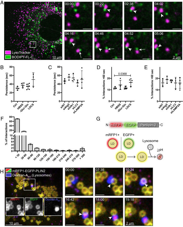
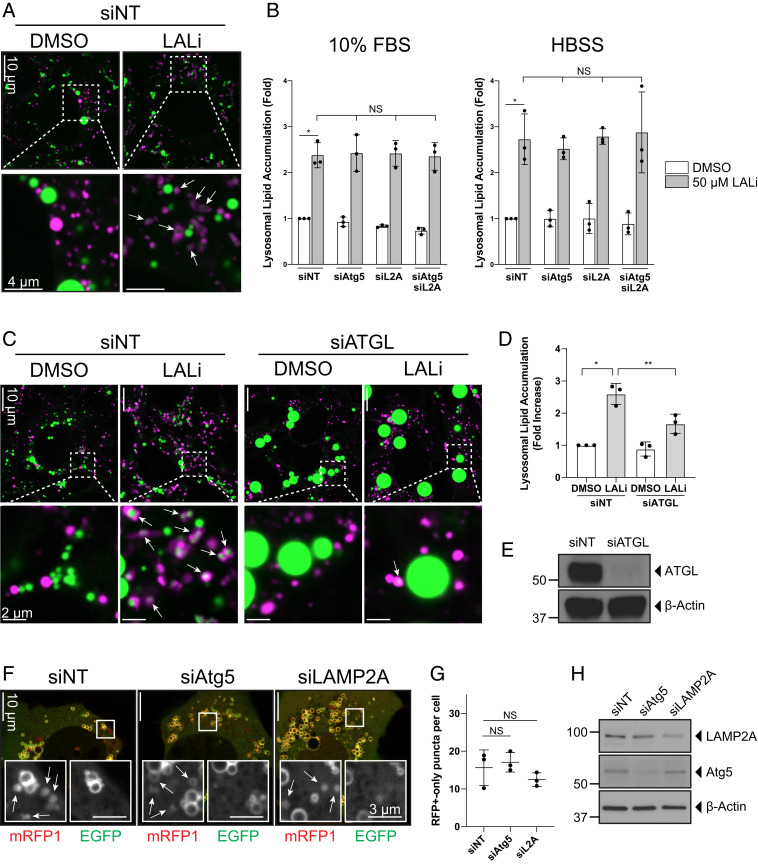
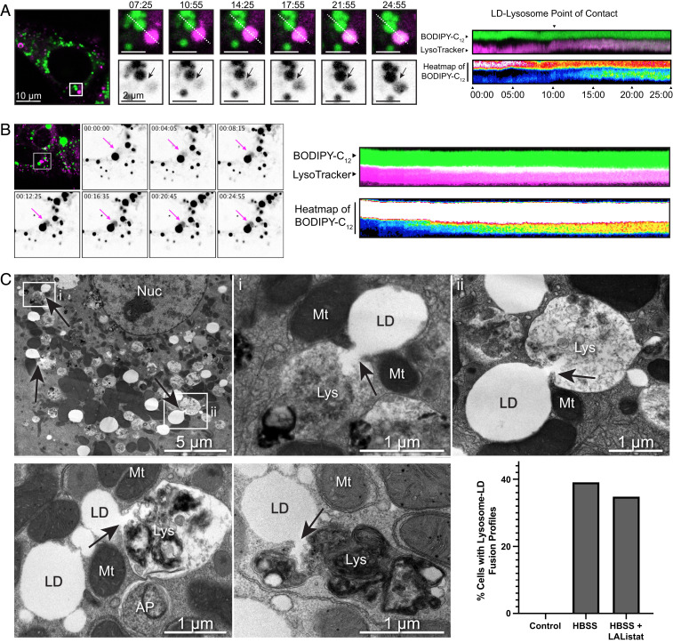
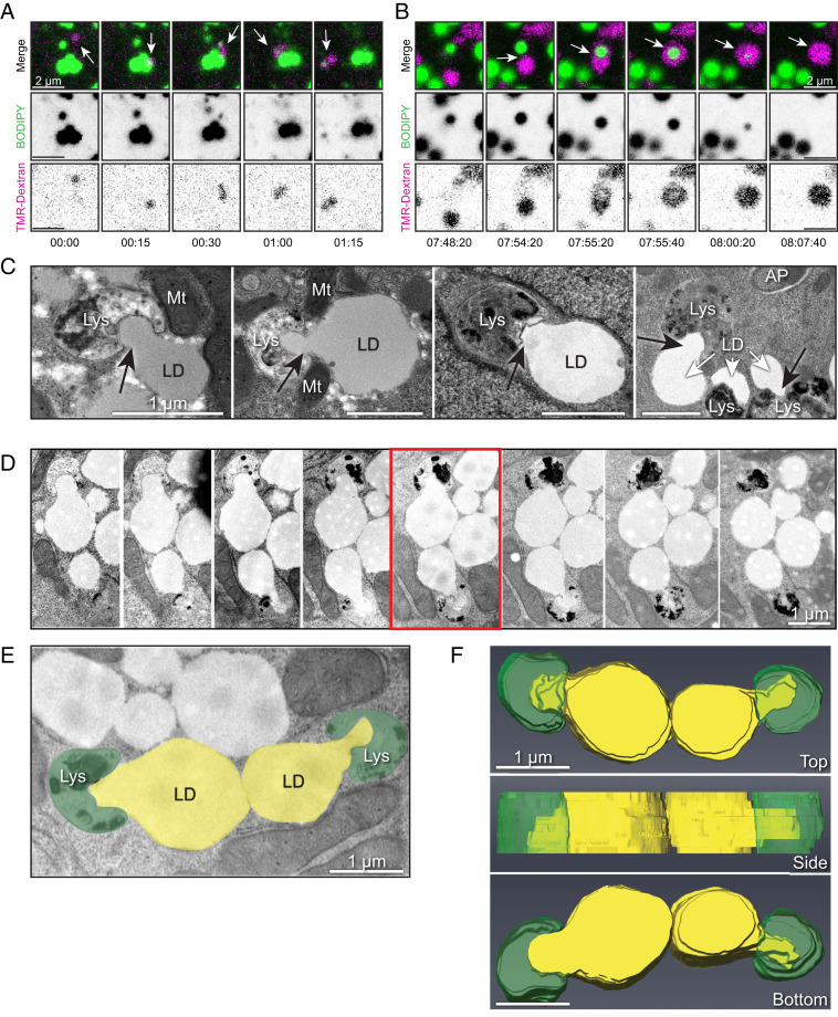
Hepatocytes metabolize energy-rich cytoplasmic lipid droplets (LDs) in the lysosome-directed process of autophagy. An organelle-selective form of this process (macrolipophagy) results in the engulfment of LDs within double-membrane delimited structures (autophagosomes) before lysosomal fusion. Whether this is an exclusive autophagic mechanism used by hepatocytes to catabolize LDs is unclear. It is also unknown whether lysosomes alone might be sufficient to mediate LD turnover in the absence of an autophagosomal intermediate. We performed live-cell microscopy of hepatocytes to monitor the dynamic interactions between lysosomes and LDs in real-time. We additionally used a fluorescent variant of the LD-specific protein (PLIN2) that exhibits altered fluorescence in response to LD interactions with the lysosome. We find that mammalian lysosomes and LDs undergo interactions during which proteins and lipids can be transferred from LDs directly into lysosomes. Electron microscopy (EM) of primary hepatocytes or hepatocyte-derived cell lines supports the existence of these interactions. It reveals a dramatic process whereby the lipid contents of the LD can be "extruded" directly into the lysosomal lumen under nutrient-limited conditions. Significantly, these interactions are not affected by perturbations to crucial components of the canonical macroautophagy machinery and can occur in the absence of double-membrane lipoautophagosomes. These findings implicate the existence of an autophagic mechanism used by mammalian cells for the direct transfer of LD components into the lysosome for breakdown. This process further emphasizes the critical role of lysosomes in hepatic LD catabolism and provides insights into the mechanisms underlying lipid homeostasis in the liver.
肝细胞通过溶酶体定向的自噬过程代谢富含细胞质脂滴 (LDs) 的能量。这种过程的一种细胞器选择性形式(巨脂噬)导致 LDs 在溶酶体融合之前被双层膜限定的结构(自噬体)吞噬。这是否是肝细胞用于分解 LDs 的一种独特的自噬机制尚不清楚。此外,在没有自噬体中间产物的情况下,单独的溶酶体是否足以介导 LD 的周转也不清楚。我们对肝细胞进行了活细胞显微镜检查,以实时监测溶酶体和 LDs 之间的动态相互作用。我们还使用了 LD 特异性蛋白 (PLIN2) 的荧光变体,该变体在与溶酶体相互作用时表现出荧光变化。我们发现哺乳动物溶酶体和 LDs 之间发生相互作用,在此过程中蛋白质和脂质可以从 LDs 直接转移到溶酶体中。对原代肝细胞或肝细胞衍生的细胞系进行的电子显微镜 (EM) 支持这些相互作用的存在。它揭示了一个戏剧性的过程,即在营养有限的条件下,LD 的脂质内容物可以“挤出”直接进入溶酶体腔。重要的是,这些相互作用不受经典巨自噬机制关键成分的干扰,并且可以在没有双层膜脂自噬体的情况下发生。这些发现表明存在一种哺乳动物细胞用于将 LD 成分直接转移到溶酶体进行分解的自噬机制。这个过程进一步强调了溶酶体在肝脏 LD 分解代谢中的关键作用,并为肝脏脂质稳态的机制提供了新的见解。