• 文献检索
  • 文档翻译
  • 深度研究
  • 学术资讯
  • Suppr Zotero 插件Zotero 插件
  • 邀请有礼
  • 套餐&价格
  • 历史记录
应用&插件
Suppr Zotero 插件Zotero 插件浏览器插件Mac 客户端Windows 客户端微信小程序
定价
高级版会员购买积分包购买API积分包
服务
文献检索文档翻译深度研究API 文档MCP 服务
关于我们
关于 Suppr公司介绍联系我们用户协议隐私条款
关注我们

Suppr 超能文献

核心技术专利:CN118964589B侵权必究
粤ICP备2023148730 号-1Suppr @ 2026

文献检索

告别复杂PubMed语法,用中文像聊天一样搜索,搜遍4000万医学文献。AI智能推荐,让科研检索更轻松。

立即免费搜索

文件翻译

保留排版,准确专业,支持PDF/Word/PPT等文件格式,支持 12+语言互译。

免费翻译文档

深度研究

AI帮你快速写综述,25分钟生成高质量综述,智能提取关键信息,辅助科研写作。

立即免费体验

线粒体蛋白加合物和超氧化物的产生是乙酰氨基酚过量后小鼠细胞溶质中 c-jun N-末端激酶早期激活的前提条件。

Mitochondrial protein adduct and superoxide generation are prerequisites for early activation of c-jun N-terminal kinase within the cytosol after an acetaminophen overdose in mice.

机构信息

Department of Pharmacology, Toxicology and Therapeutics, University of Kansas Medical Center, Kansas City, KS, 66160, USA.

Department of Pharmacology, Toxicology and Therapeutics, University of Kansas Medical Center, Kansas City, KS, 66160, USA.

出版信息

Toxicol Lett. 2021 Mar 1;338:21-31. doi: 10.1016/j.toxlet.2020.12.005. Epub 2020 Dec 5.

DOI:10.1016/j.toxlet.2020.12.005
PMID:33290831
原文链接:https://pmc.ncbi.nlm.nih.gov/articles/PMC7852579/
Abstract

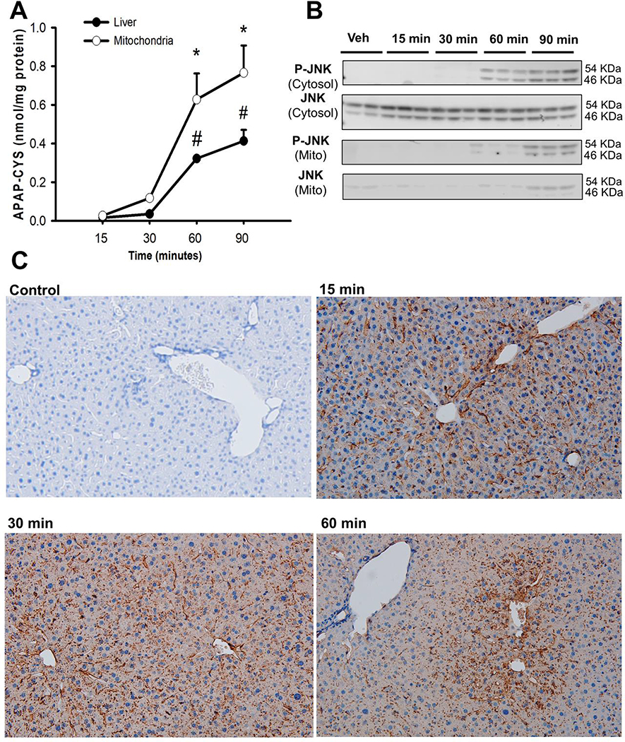
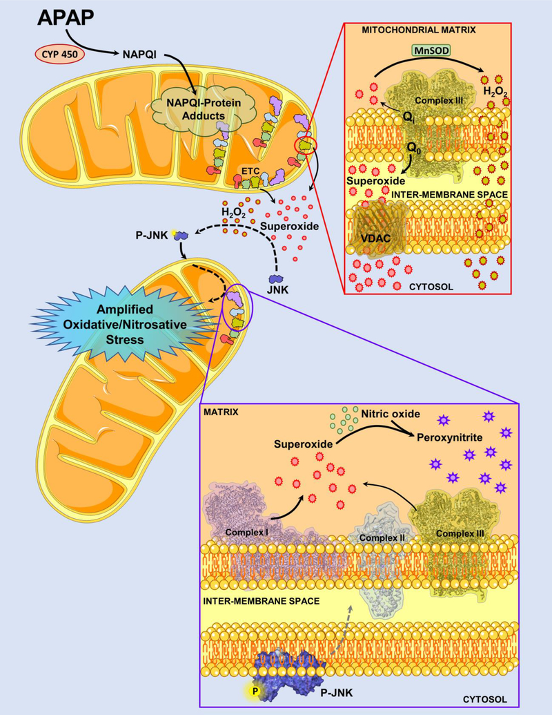
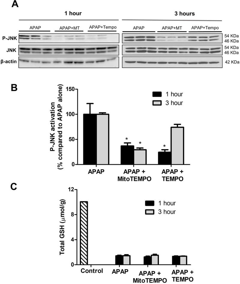
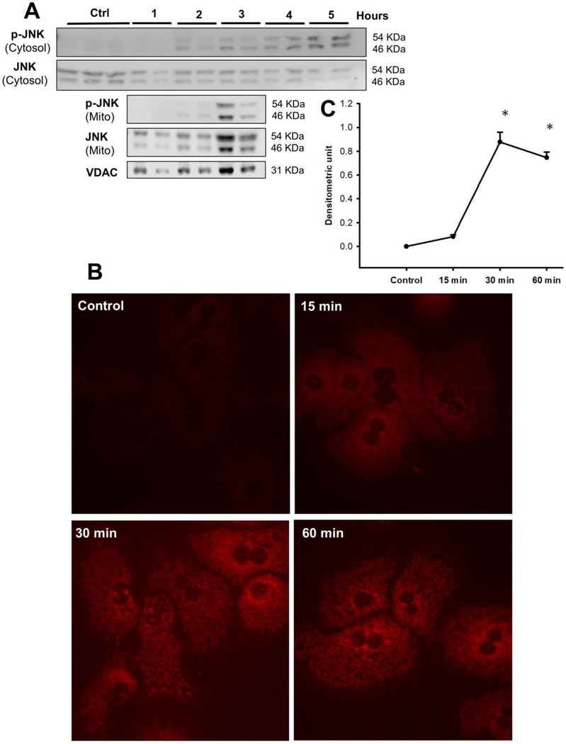
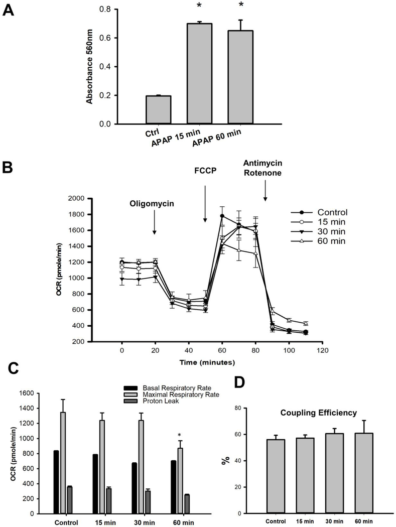
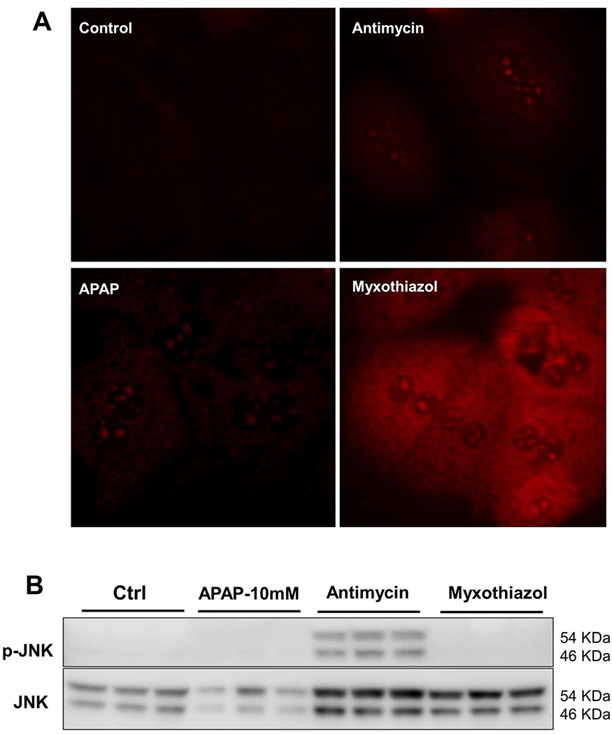
Acetaminophen (APAP) overdose is the most common cause of acute liver failure in the United States and formation of APAP-protein adducts, mitochondrial oxidant stress and activation of the mitogen activated protein (MAP) kinase c-jun N-terminal kinase (JNK) are critical for APAP-induced cell death. However, direct evidence linking these mechanistic features are lacking and were investigated by examining the early temporal course of these changes in mice after 300 mg/kg APAP. Protein adducts were detectable in the liver (0.05-0.1 nmol/mg protein) by 15 and 30 min after APAP, which increased (>500 %) selectively in mitochondria by 60 min. Cytosolic JNK activation was only evident at 60 min, and was significantly attenuated by scavenging superoxide specifically in the cytosol by TEMPO treatment. Treatment of mouse hepatocytes with APAP revealed mitochondrial superoxide generation within 15 min, accompanied by hydrogen peroxide production without change in mitochondrial respiratory function. The oxidant stress preceded JNK activation and its mitochondrial translocation. Inhibitor studies identified the putative source of mitochondrial superoxide as complex III, which released superoxide towards the intermembrane space after APAP resulting in activation of JNK in the cytosol. Our studies provide direct evidence of mechanisms involved in mitochondrial superoxide generation after NAPQI-adduct formation and its activation of the MAP kinase cascade in the cytosol, which are critical features of APAP hepatotoxicity.

摘要

对乙酰氨基酚(APAP)过量是美国急性肝衰竭的最常见原因,形成 APAP-蛋白加合物、线粒体氧化剂应激和丝裂原激活蛋白(MAP)激酶 c-jun N-末端激酶(JNK)的激活对于 APAP 诱导的细胞死亡至关重要。然而,缺乏将这些机制特征联系起来的直接证据,并通过检查 APAP 后 300mg/kg 的小鼠中这些变化的早期时间过程来研究这些证据。在 APAP 后 15 和 30 分钟,肝中可检测到蛋白加合物(0.05-0.1nmol/mg 蛋白),并在 60 分钟时选择性增加(>500%),仅在 60 分钟时可见细胞质 JNK 激活,并且通过 TEMPO 处理特异性清除细胞质中超氧阴离子显著减弱。用 APAP 处理小鼠肝细胞可在 15 分钟内产生线粒体超氧阴离子,同时伴有过氧化氢产生,而线粒体呼吸功能没有改变。氧化应激发生在 JNK 激活及其线粒体易位之前。抑制剂研究确定了线粒体超氧阴离子的潜在来源是复合物 III,APAP 后复合物 III 向膜间空间释放超氧阴离子,导致细胞质中 JNK 的激活。我们的研究提供了直接证据,证明了 NAPQI 加合物形成后线粒体超氧阴离子生成及其在细胞质中 MAP 激酶级联的激活的机制,这是 APAP 肝毒性的关键特征。

https://cdn.ncbi.nlm.nih.gov/pmc/blobs/8afe/7852579/d5a24bf8659d/nihms-1652496-f0006.jpg
https://cdn.ncbi.nlm.nih.gov/pmc/blobs/8afe/7852579/3b9b9c882eb6/nihms-1652496-f0001.jpg
https://cdn.ncbi.nlm.nih.gov/pmc/blobs/8afe/7852579/9965fa198da5/nihms-1652496-f0002.jpg
https://cdn.ncbi.nlm.nih.gov/pmc/blobs/8afe/7852579/e74ed5c71210/nihms-1652496-f0003.jpg
https://cdn.ncbi.nlm.nih.gov/pmc/blobs/8afe/7852579/dc6b154fcef5/nihms-1652496-f0004.jpg
https://cdn.ncbi.nlm.nih.gov/pmc/blobs/8afe/7852579/223f0ffbeb29/nihms-1652496-f0005.jpg
https://cdn.ncbi.nlm.nih.gov/pmc/blobs/8afe/7852579/d5a24bf8659d/nihms-1652496-f0006.jpg

相似文献

1
Mitochondrial protein adduct and superoxide generation are prerequisites for early activation of c-jun N-terminal kinase within the cytosol after an acetaminophen overdose in mice.线粒体蛋白加合物和超氧化物的产生是乙酰氨基酚过量后小鼠细胞溶质中 c-jun N-末端激酶早期激活的前提条件。
Toxicol Lett. 2021 Mar 1;338:21-31. doi: 10.1016/j.toxlet.2020.12.005. Epub 2020 Dec 5.
2
Editor's Highlight: Metformin Protects Against Acetaminophen Hepatotoxicity by Attenuation of Mitochondrial Oxidant Stress and Dysfunction.编辑亮点:二甲双胍通过减轻线粒体氧化应激和功能障碍来预防对乙酰氨基酚肝毒性。
Toxicol Sci. 2016 Dec;154(2):214-226. doi: 10.1093/toxsci/kfw158. Epub 2016 Aug 25.
3
Low Dose Acetaminophen Induces Reversible Mitochondrial Dysfunction Associated with Transient c-Jun N-Terminal Kinase Activation in Mouse Liver.低剂量对乙酰氨基酚诱导小鼠肝脏中与瞬时c-Jun氨基末端激酶激活相关的可逆性线粒体功能障碍。
Toxicol Sci. 2016 Mar;150(1):204-15. doi: 10.1093/toxsci/kfv319. Epub 2015 Dec 31.
4
Purinergic receptor antagonist A438079 protects against acetaminophen-induced liver injury by inhibiting p450 isoenzymes, not by inflammasome activation.嘌呤能受体拮抗剂 A438079 通过抑制细胞色素 P450 同工酶而不是通过炎症小体激活来防止对乙酰氨基酚引起的肝损伤。
Toxicol Sci. 2013 Jan;131(1):325-35. doi: 10.1093/toxsci/kfs283. Epub 2012 Sep 17.
5
Deletion of apoptosis signal-regulating kinase 1 attenuates acetaminophen-induced liver injury by inhibiting c-Jun N-terminal kinase activation.凋亡信号调节激酶1的缺失通过抑制c-Jun氨基末端激酶激活减轻对乙酰氨基酚诱导的肝损伤。
Gastroenterology. 2008 Oct;135(4):1311-21. doi: 10.1053/j.gastro.2008.07.006. Epub 2008 Jul 9.
6
The impact of partial manganese superoxide dismutase (SOD2)-deficiency on mitochondrial oxidant stress, DNA fragmentation and liver injury during acetaminophen hepatotoxicity.部分锰超氧化物歧化酶 (SOD2) 缺乏对乙酰氨基酚肝毒性过程中线粒体氧化剂应激、DNA 片段化和肝损伤的影响。
Toxicol Appl Pharmacol. 2011 Mar 15;251(3):226-33. doi: 10.1016/j.taap.2011.01.004. Epub 2011 Jan 15.
7
Mechanisms of acetaminophen-induced cell death in primary human hepatocytes.对乙酰氨基酚诱导原代人肝细胞死亡的机制
Toxicol Appl Pharmacol. 2014 Sep 15;279(3):266-274. doi: 10.1016/j.taap.2014.05.010. Epub 2014 Jun 3.
8
Mitochondrial protein adducts formation and mitochondrial dysfunction during N-acetyl-m-aminophenol (AMAP)-induced hepatotoxicity in primary human hepatocytes.N-乙酰间氨基酚(AMAP)诱导原代人肝细胞发生肝毒性过程中,线粒体蛋白加合物的形成及线粒体功能障碍。
Toxicol Appl Pharmacol. 2015 Dec 1;289(2):213-22. doi: 10.1016/j.taap.2015.09.022. Epub 2015 Sep 30.
9
Silencing glycogen synthase kinase-3beta inhibits acetaminophen hepatotoxicity and attenuates JNK activation and loss of glutamate cysteine ligase and myeloid cell leukemia sequence 1.沉默糖原合酶激酶-3β可抑制对乙酰氨基酚肝毒性,并减弱 JNK 激活以及谷氨酸半胱氨酸连接酶和髓样细胞白血病序列 1 的丢失。
J Biol Chem. 2010 Mar 12;285(11):8244-55. doi: 10.1074/jbc.M109.054999. Epub 2010 Jan 8.
10
c-Jun N-terminal kinase plays a major role in murine acetaminophen hepatotoxicity.c-Jun氨基末端激酶在小鼠对乙酰氨基酚肝毒性中起主要作用。
Gastroenterology. 2006 Jul;131(1):165-78. doi: 10.1053/j.gastro.2006.03.045.

引用本文的文献

1
CpG-oligodeoxynucleotides challenged macrophages ameliorate acetaminophen induced liver injury by activating TLR9/IRG1/itaconate metabolic pathway.经CpG寡脱氧核苷酸刺激的巨噬细胞通过激活TLR9/IRG1/衣康酸代谢途径改善对乙酰氨基酚诱导的肝损伤。
Mol Med. 2025 Aug 25;31(1):282. doi: 10.1186/s10020-025-01324-0.
2
Platelets mediate neutrophil infiltration and exacerbate liver injury and sinusoidal endothelial cell damage after a severe acetaminophen overdose in mice.在小鼠对乙酰氨基酚严重过量服用后,血小板介导中性粒细胞浸润,并加剧肝损伤和肝血窦内皮细胞损伤。
Arch Toxicol. 2025 Aug 16. doi: 10.1007/s00204-025-04144-7.
3
Liver sinusoidal endothelial cells secret C-X-C motif chemokine ligand 10 to promote the recruitment of invariant NKT cells in acetaminophen-induced liver injury.

本文引用的文献

1
Mitochondrial Damage and Biogenesis in Acetaminophen-induced Liver Injury.对乙酰氨基酚诱导的肝损伤中的线粒体损伤与生物发生
Liver Res. 2019 Dec;3(3-4):150-156. doi: 10.1016/j.livres.2019.10.002. Epub 2019 Nov 1.
2
A mitochondrial journey through acetaminophen hepatotoxicity.通过线粒体探索对乙酰氨基酚的肝毒性。
Food Chem Toxicol. 2020 Jun;140:111282. doi: 10.1016/j.fct.2020.111282. Epub 2020 Mar 21.
3
Acetaminophen hepatotoxicity: A mitochondrial perspective.对乙酰氨基酚肝毒性:线粒体视角
肝窦内皮细胞分泌C-X-C基序趋化因子配体10,以促进对乙酰氨基酚诱导的肝损伤中恒定自然杀伤T细胞的募集。
Sci China Life Sci. 2025 Jul 8. doi: 10.1007/s11427-025-2942-8.
4
The multiple mechanisms and modes of cell death after acetaminophen overdose.对乙酰氨基酚过量服用后细胞死亡的多种机制和模式。
Explor Dig Dis. 2025;4. doi: 10.37349/edd.2025.100569. Epub 2025 Apr 7.
5
Hepatocyte cellular repressor of E1A-stimulated genes 1 protects against acetaminophen-induced liver injury by promoting autophagy.E1A 刺激基因 1 的肝细胞细胞抑制因子 1 通过促进自噬来保护机体免受对乙酰氨基酚诱导的肝损伤。
Acta Pharmacol Sin. 2025 Mar 25. doi: 10.1038/s41401-025-01532-8.
6
The potential of curcumin in mitigating acetaminophen-induced liver damage.姜黄素在减轻对乙酰氨基酚诱导的肝损伤方面的潜力。
Naunyn Schmiedebergs Arch Pharmacol. 2025 Feb 26. doi: 10.1007/s00210-025-03907-4.
7
Mixed lineage kinase domain-like protein deficiency exacerbates early injury in a mouse model of acetaminophen hepatotoxicity.混合谱系激酶结构域样蛋白缺陷加剧对乙酰氨基酚肝毒性小鼠模型的早期损伤。
Toxicol Sci. 2025 May 1;205(1):220-232. doi: 10.1093/toxsci/kfaf022.
8
Ferroptosis and Intrinsic Drug-induced Liver Injury by Acetaminophen and Other Drugs: A Critical Evaluation and Historical Perspective.铁死亡与对乙酰氨基酚及其他药物引起的内在性药物性肝损伤:批判性评估与历史视角
J Clin Transl Hepatol. 2024 Dec 28;12(12):1057-1066. doi: 10.14218/JCTH.2024.00324. Epub 2024 Oct 29.
9
Role of Mitochondrial Iron Uptake in Acetaminophen Hepatotoxicity.线粒体铁摄取在对乙酰氨基酚肝毒性中的作用。
Livers. 2024 Sep;4(3):333-351. doi: 10.3390/livers4030024. Epub 2024 Jul 30.
10
Limitations of acetaminophen as a reference hepatotoxin for the evaluation of in vitro liver models.对乙酰氨基酚作为评估体外肝模型的参考肝毒素的局限性。
Toxicol Sci. 2025 Jan 1;203(1):35-40. doi: 10.1093/toxsci/kfae133.
Adv Pharmacol. 2019;85:195-219. doi: 10.1016/bs.apha.2019.01.007. Epub 2019 Feb 21.
4
Mitochondrial electron transport chain, ROS generation and uncoupling (Review).线粒体电子传递链、ROS 生成和解偶联(综述)。
Int J Mol Med. 2019 Jul;44(1):3-15. doi: 10.3892/ijmm.2019.4188. Epub 2019 May 8.
5
Acetaminophen Hepatotoxicity.对乙酰氨基酚肝毒性。
Semin Liver Dis. 2019 May;39(2):221-234. doi: 10.1055/s-0039-1679919. Epub 2019 Mar 8.
6
Mito-tempo protects against acute liver injury but induces limited secondary apoptosis during the late phase of acetaminophen hepatotoxicity.Mito-tempo 可预防急性肝损伤,但在对乙酰氨基酚肝毒性的晚期阶段会引起有限的继发性细胞凋亡。
Arch Toxicol. 2019 Jan;93(1):163-178. doi: 10.1007/s00204-018-2331-8. Epub 2018 Oct 15.
7
Acetaminophen-induced Acute Liver Failure Is More Common and More Severe in Women.对乙酰氨基酚导致的急性肝衰竭在女性中更为常见且更严重。
Clin Gastroenterol Hepatol. 2018 Jun;16(6):936-946. doi: 10.1016/j.cgh.2017.11.042. Epub 2017 Dec 2.
8
Acetaminophen Toxicity: Novel Insights Into Mechanisms and Future Perspectives.对乙酰氨基酚毒性:机制的新见解与未来展望
Gene Expr. 2018 Mar 21;18(1):19-30. doi: 10.3727/105221617X15084371374138. Epub 2017 Oct 20.
9
A radical shift in perspective: mitochondria as regulators of reactive oxygen species.视角的彻底转变:线粒体作为活性氧的调节者。
J Exp Biol. 2017 Apr 1;220(Pt 7):1170-1180. doi: 10.1242/jeb.132142.
10
Differential susceptibility to acetaminophen-induced liver injury in sub-strains of C57BL/6 mice: 6N versus 6J.C57BL/6小鼠亚系对乙酰氨基酚诱导的肝损伤的易感性差异:6N与6J的比较。
Food Chem Toxicol. 2016 Dec;98(Pt B):107-118. doi: 10.1016/j.fct.2016.10.021. Epub 2016 Oct 20.