Department of Electrical Engineering, Eindhoven University of Technology, 5600 MB Eindhoven, The Netherlands.
Institute for Applied Microelectronics (IUMA), University of Las Palmas de Gran Canaria (ULPGC), 35017 Las Palmas de Gran Canaria, Spain.
Sensors (Basel). 2020 Dec 5;20(23):6955. doi: 10.3390/s20236955.
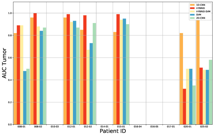
The primary treatment for malignant brain tumors is surgical resection. While gross total resection improves the prognosis, a supratotal resection may result in neurological deficits. On the other hand, accurate intraoperative identification of the tumor boundaries may be very difficult, resulting in subtotal resections. Histological examination of biopsies can be used repeatedly to help achieve gross total resection but this is not practically feasible due to the turn-around time of the tissue analysis. Therefore, intraoperative techniques to recognize tissue types are investigated to expedite the clinical workflow for tumor resection and improve outcome by aiding in the identification and removal of the malignant lesion. Hyperspectral imaging (HSI) is an optical imaging technique with the power of extracting additional information from the imaged tissue. Because HSI images cannot be visually assessed by human observers, we instead exploit artificial intelligence techniques and leverage a Convolutional Neural Network (CNN) to investigate the potential of HSI in twelve in vivo specimens. The proposed framework consists of a 3D-2D hybrid CNN-based approach to create a joint extraction of spectral and spatial information from hyperspectral images. A comparison study was conducted exploiting a 2D CNN, a 1D DNN and two conventional classification methods (SVM, and the SVM classifier combined with the 3D-2D hybrid CNN) to validate the proposed network. An overall accuracy of 80% was found when tumor, healthy tissue and blood vessels were classified, clearly outperforming the state-of-the-art approaches. These results can serve as a basis for brain tumor classification using HSI, and may open future avenues for image-guided neurosurgical applications.
恶性脑肿瘤的主要治疗方法是手术切除。虽然大体全切除可以改善预后,但超全切除可能导致神经功能缺损。另一方面,术中准确识别肿瘤边界可能非常困难,导致部分切除。活检的组织学检查可以反复使用,以帮助实现大体全切除,但由于组织分析的周转时间,这在实践中是不可行的。因此,研究了术中识别组织类型的技术,以加快肿瘤切除的临床工作流程,并通过辅助识别和切除恶性病变来改善结果。高光谱成像(HSI)是一种光学成像技术,具有从成像组织中提取附加信息的能力。由于 HSI 图像不能被人类观察者直观评估,我们转而利用人工智能技术,并利用卷积神经网络(CNN)研究 HSI 在 12 个体内标本中的潜力。所提出的框架由基于 3D-2D 混合 CNN 的方法组成,用于从高光谱图像中联合提取光谱和空间信息。进行了一项比较研究,利用二维 CNN、一维 DNN 和两种传统分类方法(SVM 和 SVM 分类器与 3D-2D 混合 CNN 相结合)来验证所提出的网络。当对肿瘤、健康组织和血管进行分类时,发现总体准确率为 80%,明显优于最先进的方法。这些结果可以为使用 HSI 进行脑肿瘤分类提供基础,并可能为图像引导神经外科应用开辟新的途径。