Department of Radiation Oncology and Molecular Radiation Sciences, Johns Hopkins University School of Medicine, Baltimore, Maryland, USA.
Division of Cardiology, Department of Medicine, Johns Hopkins University School of Medicine, Baltimore, Maryland, USA.
J Appl Clin Med Phys. 2022 Sep;23(9):e13725. doi: 10.1002/acm2.13725. Epub 2022 Jul 27.
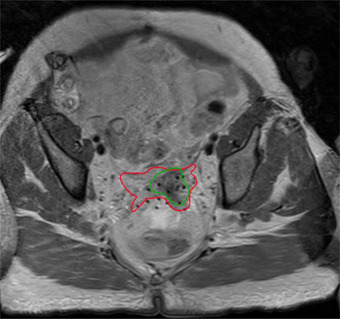
Contouring clinical target volume (CTV) from medical images is an essential step for radiotherapy (RT) planning. Magnetic resonance imaging (MRI) is used as a standard imaging modality for CTV segmentation in cervical cancer due to its superior soft-tissue contrast. However, the delineation of CTV is challenging as CTV contains microscopic extensions that are not clearly visible even in MR images, resulting in significant contour variability among radiation oncologists depending on their knowledge and experience. In this study, we propose a fully automated deep learning-based method to segment CTV from MR images.
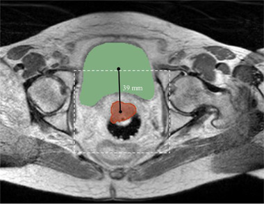
Our method begins with the bladder segmentation, from which the CTV position is estimated in the axial view. The superior-inferior CTV span is then detected using an Attention U-Net. A CTV-specific region of interest (ROI) is determined, and three-dimensional (3-D) blocks are extracted from the ROI volume. Finally, a CTV segmentation map is computed using a 3-D U-Net from the extracted 3-D blocks.
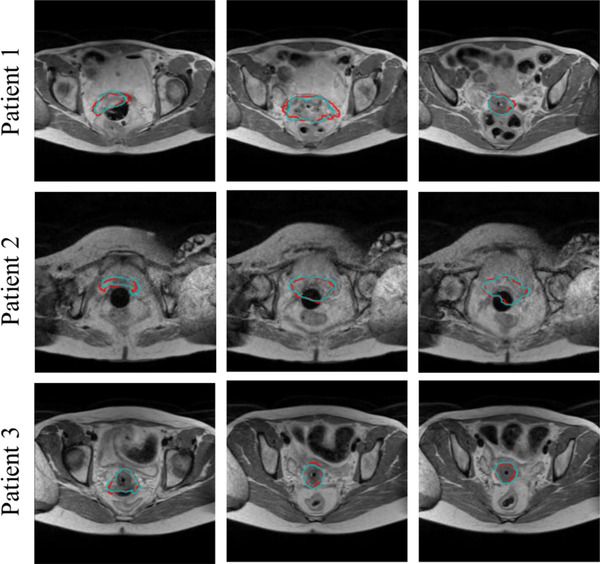
We developed and evaluated our method using 213 MRI scans obtained from 125 patients (183 for training, 30 for test). Our method achieved (mean ± SD) Dice similarity coefficient of 0.85 ± 0.03 and the 95th percentile Hausdorff distance of 3.70 ± 0.35 mm on test cases, outperforming other state-of-the-art methods significantly (p-value < 0.05). Our method also produces an uncertainty map along with the CTV segmentation by employing the Monte Carlo dropout technique to draw physician's attention to the regions with high uncertainty, where careful review and manual correction may be needed.
Experimental results show that the developed method is accurate, fast, and reproducible for contouring CTV from MRI, demonstrating its potential to assist radiation oncologists in alleviating the burden of tedious contouring for RT planning in cervical cancer.
从医学图像中勾画临床靶区(CTV)是放射治疗(RT)计划的重要步骤。由于磁共振成像(MRI)具有优越的软组织对比度,因此它被用作宫颈癌 CTV 分割的标准成像方式。然而,由于 CTV 包含即使在 MRI 图像中也无法清晰看到的微观延伸,因此 CTV 的勾画具有挑战性,这导致不同的放射肿瘤学家根据他们的知识和经验,CTV 的勾画存在很大的差异。在本研究中,我们提出了一种完全基于深度学习的自动分割 MRI 图像中 CTV 的方法。
我们的方法从膀胱分割开始,从轴向视图中估计 CTV 的位置。然后使用注意 U-Net 检测上下 CTV 跨度。确定 CTV 特定的感兴趣区域(ROI),并从 ROI 体积中提取三维(3-D)块。最后,使用从提取的 3-D 块计算的 3-D U-Net 计算 CTV 分割图。
我们使用从 125 名患者获得的 213 个 MRI 扫描(183 个用于训练,30 个用于测试)开发并评估了我们的方法。我们的方法在测试病例上实现了(平均值±标准差)Dice 相似系数为 0.85±0.03,95%Hausdorff 距离为 3.70±0.35mm,明显优于其他最先进的方法(p 值<0.05)。我们的方法还通过使用蒙特卡罗辍学技术生成 CTV 分割的不确定性图,提请医生注意高不确定性区域,从而实现了快速、准确和可重复的 CTV 勾画,需要仔细审查和手动校正。
实验结果表明,所开发的方法在从 MRI 勾画 CTV 方面准确、快速且可重复,展示了其在减轻宫颈癌 RT 计划中繁琐勾画负担方面辅助放射肿瘤学家的潜力。