Department of Radiology, Seoul National University Hospital, Jongno-gu, Seoul, Republic of Korea.
Department of Radiology, Seoul National University College of Medicine, Jongno-gu, Seoul, Republic of Korea.
PLoS One. 2020 Dec 11;15(12):e0243815. doi: 10.1371/journal.pone.0243815. eCollection 2020.
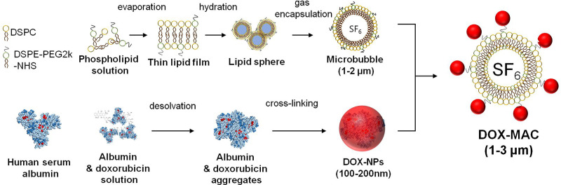
To assess the feasibility of the combined sorafenib (SOR) and doxorubicin-loaded microbubble-albumin nanoparticle complex (DOX-MAC) treatment effect in an orthotopic rat model of hepatocellular carcinoma (HCC).
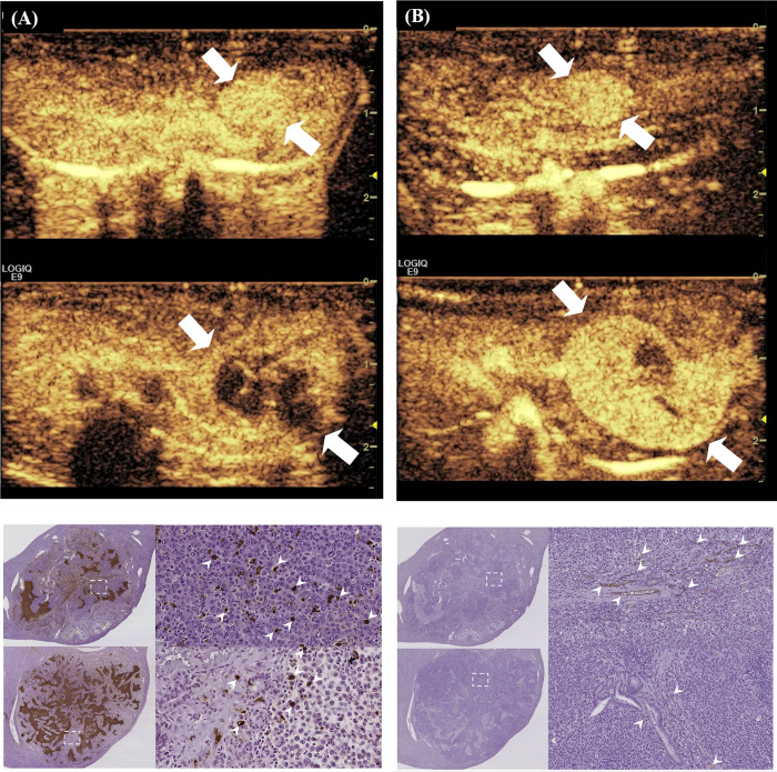
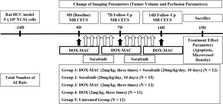
Sixty-two rats with N1-S1 hepatoma were divided into four groups according to the treatment methods, i.e. G1 (SOR and DOX-MAC; n = 12), G2 (SOR; n = 15), G3 (DOX-MAC; n = 12), G4 (DOX; n = 11), and G5 (normal saline; n = 12). We performed the theragnostic, contrast-enhanced ultrasound examination and treatment at the baseline, one-week, and two-weeks. Tumor volume and perfusion parameters were compared at each time point and the differences between all of the groups over time were analyzed using repeated measures ANOVA. We also analyzed the apoptotic index and microvessel density (MVD) per each tumor specimen in all of the groups.
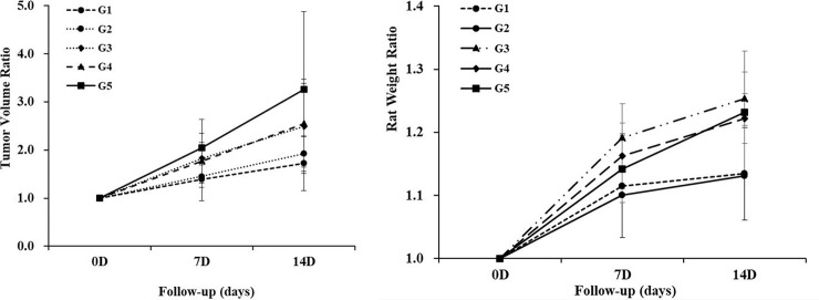
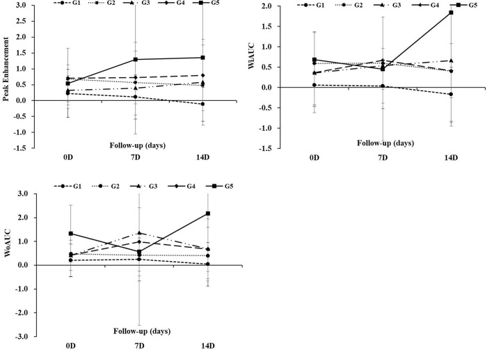
The tumors increased from the beginning in all of the groups to the final follow-up, whereas the tumor growth in the G1 group and the G2 group was inhibited during the treatment period compared to the baseline tumor volume (P = 0.016 and P = 0.031). The G1 group resulted in tumor growth inhibition compared to the control group (P = 0.008). The G1 group showed that the peak enhancement and wash-in area under the curve were lower than that of the G4 group (P = 0.010 and 0.022). However, there was no difference in perfusion parameters in the other treated group compared to control group. The MVD of the G1 group tumor was lower than that of the G4 group (P = .016).
Our results suggest that the combination therapy of SOR and DOX-MAC can cause inhibition of tumor growth after treatment and that this therapy can be adequately monitored using the theragnostic DOX-MAC agent.
评估索拉非尼(SOR)和多柔比星载微泡白蛋白纳米复合物(DOX-MAC)联合治疗在原位大鼠肝癌(HCC)模型中的可行性。
62 只 N1-S1 肝癌大鼠按治疗方法分为 4 组,即 G1 组(SOR 和 DOX-MAC;n = 12)、G2 组(SOR;n = 15)、G3 组(DOX-MAC;n = 12)、G4 组(DOX;n = 11)和 G5 组(生理盐水;n = 12)。我们在基线、1 周和 2 周时进行了诊断和治疗的对比增强超声检查。在每个时间点比较肿瘤体积和灌注参数,并使用重复测量方差分析分析所有组随时间的差异。我们还分析了所有组的每个肿瘤标本的凋亡指数和微血管密度(MVD)。
所有组的肿瘤从开始到最终随访都在增加,而 G1 组和 G2 组在治疗期间的肿瘤生长与基线肿瘤体积相比受到抑制(P = 0.016 和 P = 0.031)。与对照组相比,G1 组导致肿瘤生长抑制(P = 0.008)。G1 组的峰值增强和灌洗曲线下面积低于 G4 组(P = 0.010 和 0.022)。然而,与对照组相比,其他治疗组的灌注参数没有差异。G1 组肿瘤的 MVD 低于 G4 组(P = 0.016)。
我们的结果表明,SOR 和 DOX-MAC 的联合治疗在治疗后可引起肿瘤生长抑制,并且这种治疗可以通过诊断性 DOX-MAC 剂充分监测。