Shanghai Eye Disease Prevention and Treatment Center, Shanghai Eye Hospital, Shanghai, China.
Department of Ophthalmology, Shanghai General Hospital, Shanghai Jiao Tong University, National Clinical Research Center for Eye Diseases, Shanghai Key Laboratory of Ocular Fundus Diseases, Shanghai Engineering Center for Visual Science and Photomedicine, Shanghai Engineering Center for Precise Diagnosis and Treatment of Eye Diseases, Shanghai, China.
Invest Ophthalmol Vis Sci. 2020 Dec 1;61(14):15. doi: 10.1167/iovs.61.14.15.
To examine the changes in choroidal thickness (ChT) after 6 months of 1% or 0.01% atropine treatment and the independent factors associated with eye elongation.
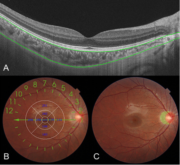
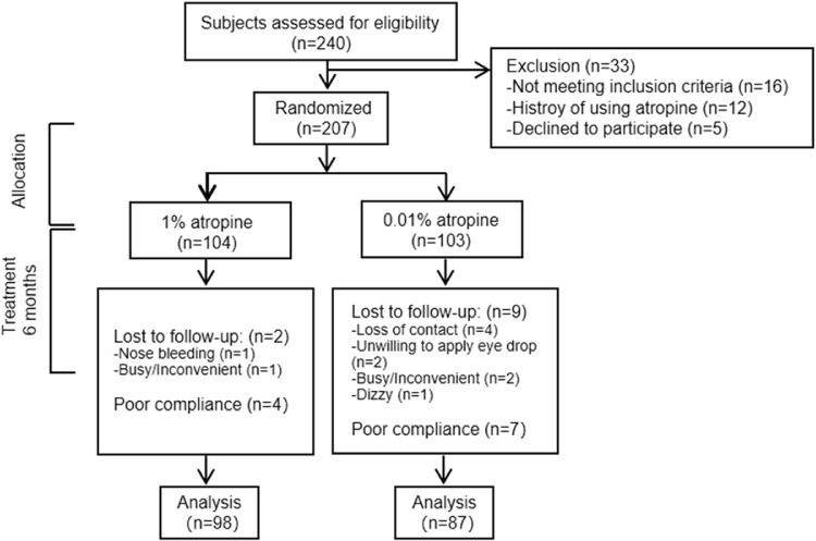
A total of 207 myopic children aged 6 to 12 years were recruited and randomly assigned to groups A and B in a ratio of 1:1. Participants in group A received 1% atropine once a day for 1 week, and then once a week for 23 weeks. Participants in group B received 0.01% atropine once a day for 6 months. ChT and internal axial length (IAL) were measured at baseline, 1 week, 3 months, and 6 months.
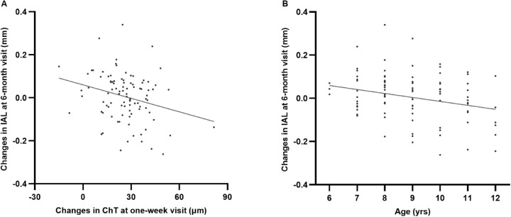
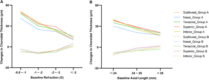
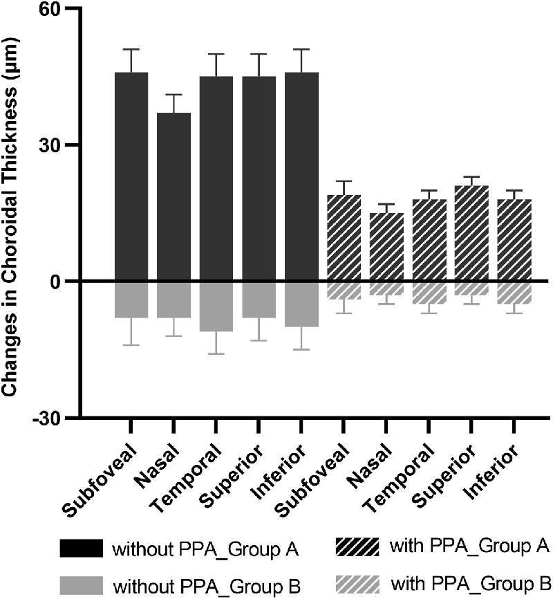
In group A, the ChT significantly increased after a 1-week loading dose of 1% atropine (26 ± 14 µm; P < 0.001) and the magnitude of increase stabilized throughout the following weekly treatment. The internal axial length did not significantly change at the 6-month visit (-0.01 ± 0.11 mm; P = 0.74). In contrast, a decreased ChT (-5 ± 17 µm; P < 0.001) and pronounced eye elongation (0.19 ± 0.12 mm; P < 0.001) were observed in group B after 6 months. Multivariable regression analysis showed that less increase in ChT at the 1-week visit (P = 0.03), younger age (P < 0.001), and presence of peripapillary atrophy (P = 0.001) were significantly associated with greater internal axial length increase over 6 months in group A.
One percent atropine could increase the ChT, whereas 0.01% atropine caused a decrease in ChT after 6 months of treatment. For participants receiving 1% atropine, the short-term increase in ChT was negatively associated with long-term eye elongation. Younger age and the presence of peripapillary atrophy were found to be risk factors for greater eye elongation.
观察 6 个月 1%或 0.01%阿托品治疗后脉络膜厚度(ChT)的变化,并探讨与眼轴延长相关的独立因素。
共招募 207 名 6 至 12 岁的近视儿童,按照 1:1 的比例随机分为 A 组和 B 组。A 组参与者每天接受 1%阿托品治疗 1 周,然后每周治疗 23 周。B 组参与者每天接受 0.01%阿托品治疗 6 个月。在基线、1 周、3 个月和 6 个月时测量 ChT 和眼内轴长(IAL)。
A 组中,1%阿托品负荷剂量治疗 1 周后 ChT 明显增加(26±14 µm;P<0.001),且在随后的每周治疗中增加幅度稳定。6 个月时 IAL 无明显变化(-0.01±0.11 mm;P=0.74)。相比之下,B 组治疗 6 个月后 ChT 减少(-5±17 µm;P<0.001),眼轴明显延长(0.19±0.12 mm;P<0.001)。多变量回归分析显示,A 组中第 1 周时 ChT 增加较少(P=0.03)、年龄较小(P<0.001)和存在视盘周围萎缩(P=0.001)与 6 个月内 IAL 增加较大显著相关。
1%阿托品可增加 ChT,而 0.01%阿托品治疗 6 个月后可导致 ChT 减少。对于接受 1%阿托品治疗的患者,短期 ChT 增加与长期眼轴延长呈负相关。年龄较小和存在视盘周围萎缩被认为是眼轴延长较大的危险因素。