• 文献检索
  • 文档翻译
  • 深度研究
  • 学术资讯
  • Suppr Zotero 插件Zotero 插件
  • 邀请有礼
  • 套餐&价格
  • 历史记录
应用&插件
Suppr Zotero 插件Zotero 插件浏览器插件Mac 客户端Windows 客户端微信小程序
定价
高级版会员购买积分包购买API积分包
服务
文献检索文档翻译深度研究API 文档MCP 服务
关于我们
关于 Suppr公司介绍联系我们用户协议隐私条款
关注我们

Suppr 超能文献

核心技术专利:CN118964589B侵权必究
粤ICP备2023148730 号-1Suppr @ 2026

文献检索

告别复杂PubMed语法,用中文像聊天一样搜索,搜遍4000万医学文献。AI智能推荐,让科研检索更轻松。

立即免费搜索

文件翻译

保留排版,准确专业,支持PDF/Word/PPT等文件格式,支持 12+语言互译。

免费翻译文档

深度研究

AI帮你快速写综述,25分钟生成高质量综述,智能提取关键信息,辅助科研写作。

立即免费体验

肾小管上皮细胞来源的外泌体微小RNA-374b-5p促进M1巨噬细胞活化并加重肾脏缺血/再灌注损伤。

Exosomal MicroRNA-374b-5p From Tubular Epithelial Cells Promoted M1 Macrophages Activation and Worsened Renal Ischemia/Reperfusion Injury.

作者信息

Ding Chenguang, Zheng Jin, Wang Bo, Li Yang, Xiang Heli, Dou Meng, Qiao Yuxi, Tian Puxun, Ding Xiaoming, Xue Wujun

机构信息

Department of Kidney Transplantation, Nephropathy Hospital, The First Affiliated Hospital, Xi'an Jiaotong University, Xi'an, China.

Institute of Organ Transplantation, The First Affiliated Hospital, Xi'an Jiaotong University, Xi'an, China.

出版信息

Front Cell Dev Biol. 2020 Nov 26;8:587693. doi: 10.3389/fcell.2020.587693. eCollection 2020.

DOI:10.3389/fcell.2020.587693
PMID:33324643
原文链接:https://pmc.ncbi.nlm.nih.gov/articles/PMC7726230/
Abstract

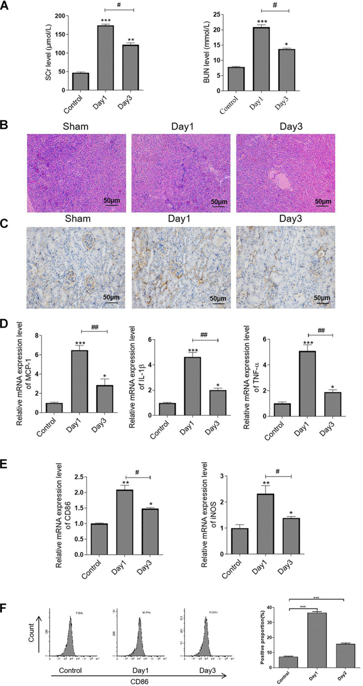
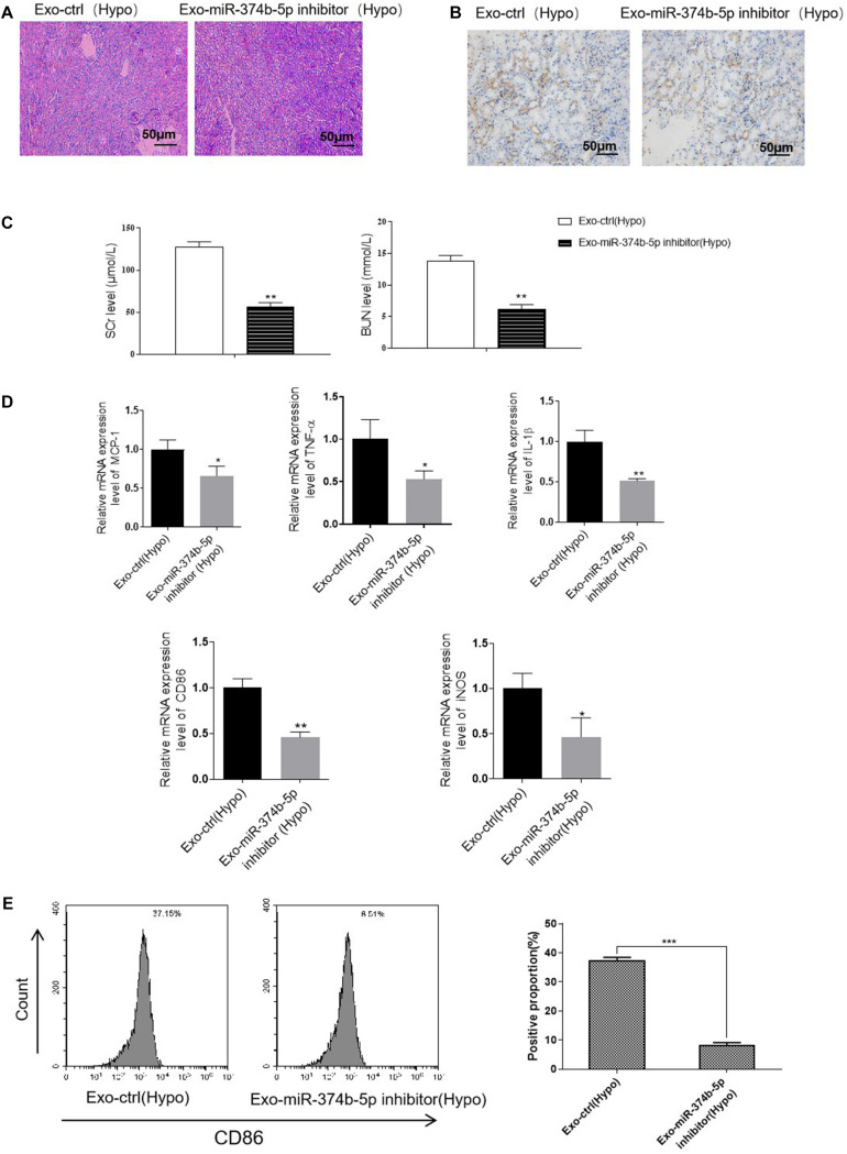
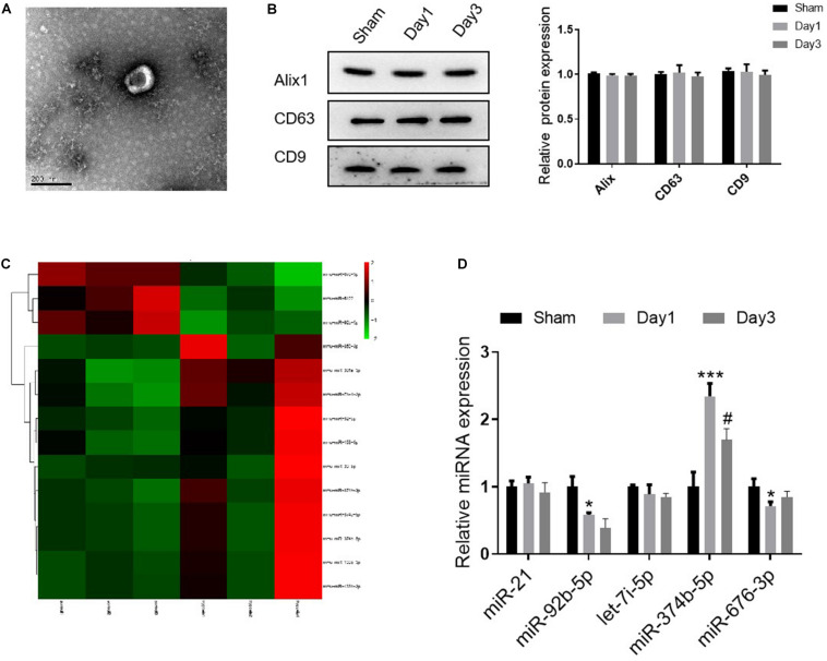
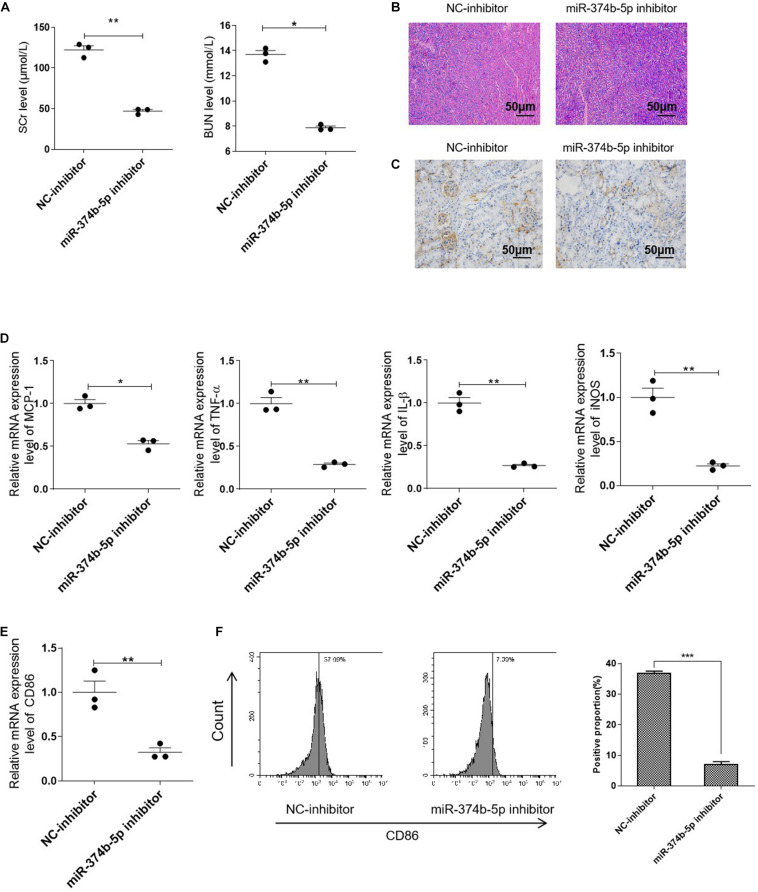
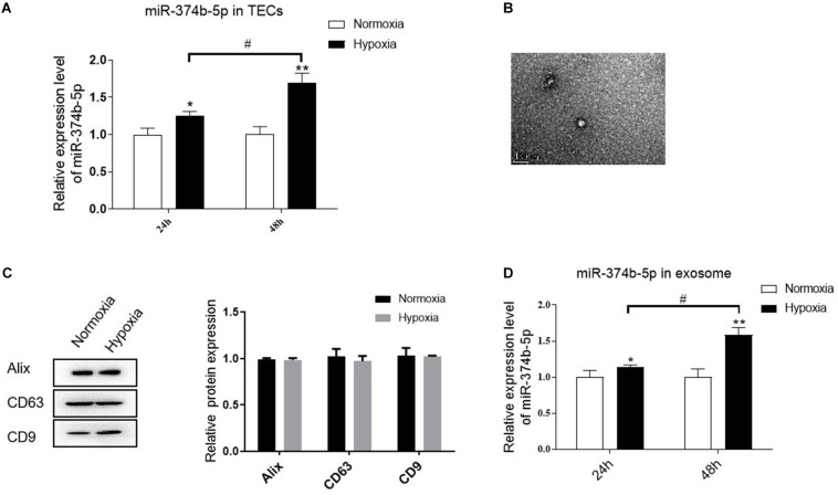
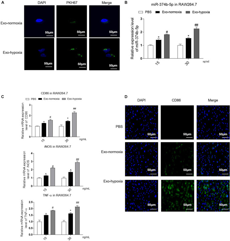
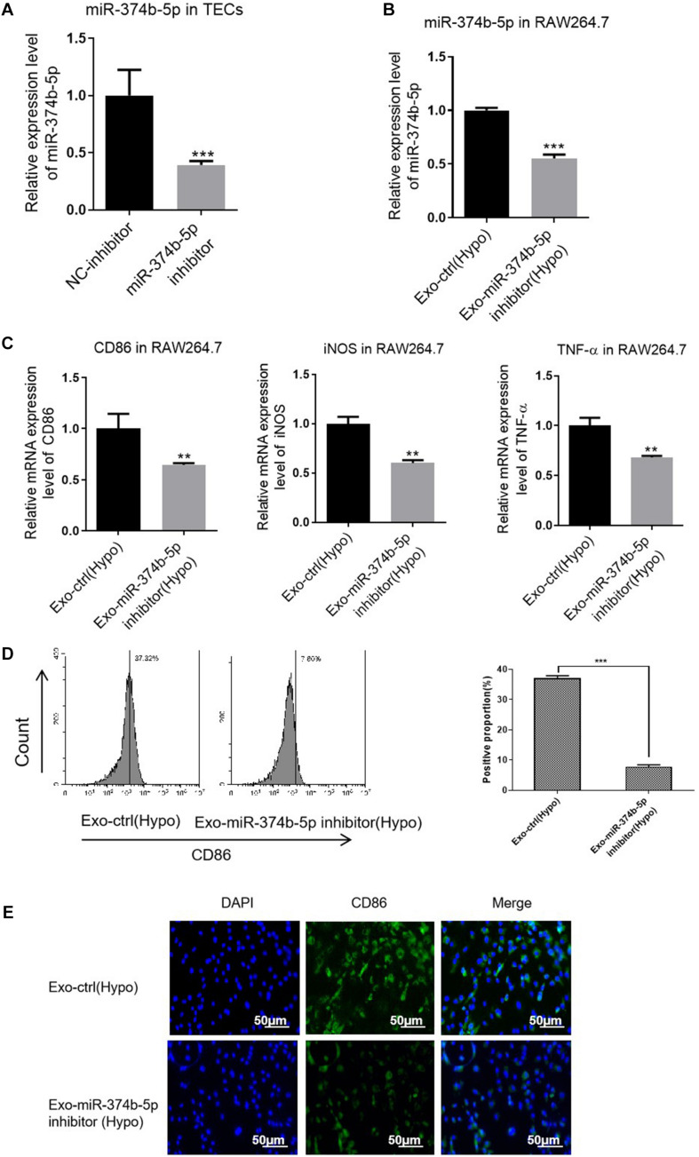
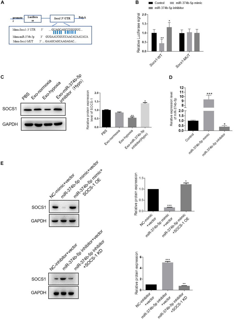
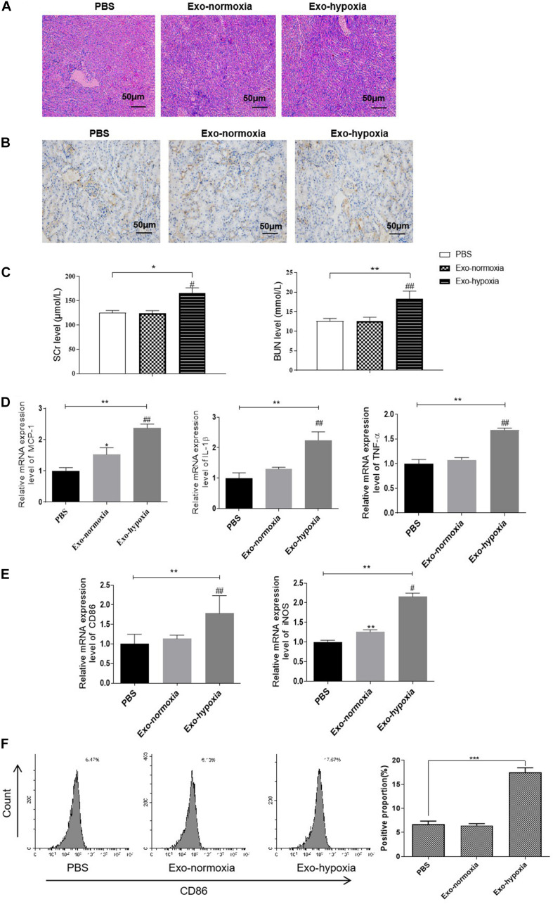
Tubular epithelial cells (TECs) represent the primary site of renal ischemia/reperfusion injury (RIRI). However, whether the damage of TECs could drive the initiation of inflammation was unclear. Here we investigated the role of the TECs and macrophages during RIRI. Increased expression of inflammation response and activated M1 macrophage were determined in the mice model of RIRI. Moreover, we demonstrated global miRNA expression profiling of renal exosomes, and miR-374b-5p was most upregulated in these exosomes . Inhibition of miR-374b-5p in the mice upon RIR operation would alleviate the kidney injury via decreasing the production of proinflammatory cytokines and suppressing the macrophage activation. Similar results were also identified in the hypoxia-induced cell model where exosomal miR-374b-5p was dramatically upregulated. Uptake of exosomes derived from the hypoxic TECs by macrophages would trigger M1 polarization via transferring miR-374b-5p. Besides, we confirmed that miR-374b-5p could directly bind to using a dual-luciferase reporter assay. Notably, when we injected the miR-374b-5p-enriched exosomes into mice, a high-level inflammatory response and M1 macrophage activation were performed. Our studies demonstrated that exosomal miR-374b-5p played an essential role in the communication between injured TECs and macrophages, resulting in the M1 macrophage activation during RIRI. The blockage of the release of such exosomes may serve as a new therapeutic strategy for RIRI.

摘要

肾小管上皮细胞(TECs)是肾脏缺血/再灌注损伤(RIRI)的主要发生部位。然而,TECs的损伤是否会引发炎症反应尚不清楚。在此,我们研究了TECs和巨噬细胞在RIRI过程中的作用。在RIRI小鼠模型中,检测到炎症反应的表达增加以及M1巨噬细胞的激活。此外,我们对肾外泌体的整体miRNA表达谱进行了分析,发现miR-374b-5p在这些外泌体中上调最为明显。在RIR手术后对小鼠体内的miR-374b-5p进行抑制,可通过减少促炎细胞因子的产生和抑制巨噬细胞激活来减轻肾脏损伤。在缺氧诱导的细胞模型中也得到了类似的结果,其中外泌体miR-374b-5p显著上调。巨噬细胞摄取缺氧TECs来源的外泌体可通过转运miR-374b-5p触发M1极化。此外,我们通过双荧光素酶报告基因实验证实miR-374b-5p可以直接结合 。值得注意的是,当我们将富含miR-374b-5p的外泌体注射到小鼠体内时,会引发高水平的炎症反应和M1巨噬细胞激活。我们的研究表明,外泌体miR-374b-5p在受损TECs与巨噬细胞之间的通讯中起重要作用,导致RIRI过程中M1巨噬细胞的激活。阻断此类外泌体的释放可能成为RIRI的一种新的治疗策略。

https://cdn.ncbi.nlm.nih.gov/pmc/blobs/7aeb/7726230/bc432756ef83/fcell-08-587693-g009.jpg
https://cdn.ncbi.nlm.nih.gov/pmc/blobs/7aeb/7726230/d3ca6321eccc/fcell-08-587693-g001.jpg
https://cdn.ncbi.nlm.nih.gov/pmc/blobs/7aeb/7726230/ac34b18d5a00/fcell-08-587693-g002.jpg
https://cdn.ncbi.nlm.nih.gov/pmc/blobs/7aeb/7726230/bcd03c62d798/fcell-08-587693-g003.jpg
https://cdn.ncbi.nlm.nih.gov/pmc/blobs/7aeb/7726230/2e1544c28091/fcell-08-587693-g004.jpg
https://cdn.ncbi.nlm.nih.gov/pmc/blobs/7aeb/7726230/301288235a5d/fcell-08-587693-g005.jpg
https://cdn.ncbi.nlm.nih.gov/pmc/blobs/7aeb/7726230/fbf0eddc3ca2/fcell-08-587693-g006.jpg
https://cdn.ncbi.nlm.nih.gov/pmc/blobs/7aeb/7726230/626764c2671b/fcell-08-587693-g007.jpg
https://cdn.ncbi.nlm.nih.gov/pmc/blobs/7aeb/7726230/2d1ef27b2823/fcell-08-587693-g008.jpg
https://cdn.ncbi.nlm.nih.gov/pmc/blobs/7aeb/7726230/bc432756ef83/fcell-08-587693-g009.jpg

相似文献

1
Exosomal MicroRNA-374b-5p From Tubular Epithelial Cells Promoted M1 Macrophages Activation and Worsened Renal Ischemia/Reperfusion Injury.肾小管上皮细胞来源的外泌体微小RNA-374b-5p促进M1巨噬细胞活化并加重肾脏缺血/再灌注损伤。
Front Cell Dev Biol. 2020 Nov 26;8:587693. doi: 10.3389/fcell.2020.587693. eCollection 2020.
2
Exosomal miRNA-19b-3p of tubular epithelial cells promotes M1 macrophage activation in kidney injury.肾小管上皮细胞来源的外泌体 miR-19b-3p 促进肾损伤中 M1 巨噬细胞的活化。
Cell Death Differ. 2020 Jan;27(1):210-226. doi: 10.1038/s41418-019-0349-y. Epub 2019 May 16.
3
Macrophage-derived exosomal miR-195a-5p impairs tubular epithelial cells mitochondria in acute kidney injury mice.巨噬细胞衍生的外泌体 miR-195a-5p 损害急性肾损伤小鼠肾小管上皮细胞的线粒体。
FASEB J. 2023 Jan;37(1):e22691. doi: 10.1096/fj.202200644R.
4
Ischemia and reperfusion-injured liver-derived exosomes elicit acute lung injury through miR-122-5p regulated alveolar macrophage polarization.缺血再灌注损伤的肝脏来源外泌体通过 miR-122-5p 调控的肺泡巨噬细胞极化引发急性肺损伤。
Int Immunopharmacol. 2024 Apr 20;131:111853. doi: 10.1016/j.intimp.2024.111853. Epub 2024 Mar 18.
5
Tubular cell-derived exosomal miR-150-5p contributes to renal fibrosis following unilateral ischemia-reperfusion injury by activating fibroblast and .管状细胞来源的外泌体 miR-150-5p 通过激活成纤维细胞和 ,导致单侧缺血再灌注损伤后的肾脏纤维化。
Int J Biol Sci. 2021 Sep 21;17(14):4021-4033. doi: 10.7150/ijbs.62478. eCollection 2021.
6
Inhibition of MiR-106b-5p mediated by exosomes mitigates acute kidney injury by modulating transmissible endoplasmic reticulum stress and M1 macrophage polarization.外泌体介导的MiR-106b-5p抑制通过调节可传播的内质网应激和M1巨噬细胞极化减轻急性肾损伤。
J Cell Mol Med. 2023 Oct;27(19):2876-2889. doi: 10.1111/jcmm.17848. Epub 2023 Jul 20.
7
miR-20a-5p is enriched in hypoxia-derived tubular exosomes and protects against acute tubular injury.miR-20a-5p 富集于低氧衍生的肾小管细胞外泌体中,可防止急性肾小管损伤。
Clin Sci (Lond). 2020 Aug 28;134(16):2223-2234. doi: 10.1042/CS20200288.
8
Exosomal miR-155-5p drives widespread macrophage M1 polarization in hypervirulent Klebsiella pneumoniae-induced acute lung injury via the MSK1/p38-MAPK axis.外泌体 miR-155-5p 通过 MSK1/p38-MAPK 轴驱动高毒力肺炎克雷伯菌诱导的急性肺损伤中广泛的巨噬细胞 M1 极化。
Cell Mol Biol Lett. 2023 Nov 13;28(1):92. doi: 10.1186/s11658-023-00505-1.
9
Macrophage-derived exosomes promote telomere fragility and senescence in tubular epithelial cells by delivering miR-155.巨噬细胞衍生的外泌体通过递送 miR-155 促进管状上皮细胞端粒脆弱和衰老。
Cell Commun Signal. 2024 Jul 10;22(1):357. doi: 10.1186/s12964-024-01708-5.
10
Injured tubular epithelial cells activate fibroblasts to promote kidney fibrosis through miR-150-containing exosomes.受损的管状上皮细胞通过含有 miR-150 的外泌体激活成纤维细胞,促进肾脏纤维化。
Exp Cell Res. 2020 Jul 15;392(2):112007. doi: 10.1016/j.yexcr.2020.112007. Epub 2020 Apr 18.

引用本文的文献

1
The role of macrophages in renal fibrosis and therapeutic prospects.巨噬细胞在肾纤维化中的作用及治疗前景。
PeerJ. 2025 Jul 23;13:e19769. doi: 10.7717/peerj.19769. eCollection 2025.
2
Mechanisms of Acute Kidney Injury-Chronic Kidney Disease Transition: Unraveling Maladaptive Repair and Therapeutic Opportunities.急性肾损伤-慢性肾脏病转化机制:解析适应性不良修复及治疗机遇
Biomolecules. 2025 May 29;15(6):794. doi: 10.3390/biom15060794.
3
Exosomal let-7b-5p derived from Aspergillus fumigatus-treated human corneal epithelial cells promotes M1 macrophage activation via targeting SOCS-1.

本文引用的文献

1
The biology function and biomedical applications of exosomes.外泌体的生物学功能和生物医学应用。
Science. 2020 Feb 7;367(6478). doi: 10.1126/science.aau6977.
2
Exosomes Derived From Mesenchymal Stem Cells Ameliorate Renal Ischemic-Reperfusion Injury Through Inhibiting Inflammation and Cell Apoptosis.间充质干细胞来源的外泌体通过抑制炎症和细胞凋亡减轻肾脏缺血再灌注损伤。
Front Med (Lausanne). 2019 Nov 19;6:269. doi: 10.3389/fmed.2019.00269. eCollection 2019.
3
Calcium channel Orai1 promotes lymphocyte IL-17 expression and progressive kidney injury.
源自烟曲霉处理的人角膜上皮细胞的外泌体let-7b-5p通过靶向SOCS-1促进M1巨噬细胞活化。
Front Immunol. 2025 Jun 4;16:1548802. doi: 10.3389/fimmu.2025.1548802. eCollection 2025.
4
Extracellular Vesicles in Renal Inflammatory Diseases: Revealing Mechanisms of Extracellular Vesicle-Mediated Macrophage Regulation.肾脏炎症性疾病中的细胞外囊泡:揭示细胞外囊泡介导的巨噬细胞调节机制
Int J Mol Sci. 2025 Apr 12;26(8):3646. doi: 10.3390/ijms26083646.
5
Effects and Mechanisms of Extracellular Vesicles in Different Models of Acute Kidney Injury.细胞外囊泡在不同急性肾损伤模型中的作用及机制
Stem Cells Int. 2025 Mar 24;2025:1075016. doi: 10.1155/sci/1075016. eCollection 2025.
6
Emerging Frontiers in acute kidney injury: The role of extracellular vesicles.急性肾损伤的新兴前沿领域:细胞外囊泡的作用
Bioact Mater. 2025 Feb 18;48:149-170. doi: 10.1016/j.bioactmat.2025.02.018. eCollection 2025 Jun.
7
Piezo1 aggravates ischemia/reperfusion-induced acute kidney injury by Ca-dependent calpain/HIF-1α/Notch signaling.Piezo1通过钙依赖性钙蛋白酶/HIF-1α/Notch信号通路加重缺血/再灌注诱导的急性肾损伤。
Ren Fail. 2025 Dec;47(1):2447801. doi: 10.1080/0886022X.2024.2447801. Epub 2025 Jan 8.
8
MiR-92a-3p Promotes Renal Injury and Fibrosis Through Facilitating M1 Macrophage Polarization via Targeting LIN28A.miR-92a-3p 通过靶向 LIN28A 促进 M1 巨噬细胞极化从而促进肾损伤和纤维化。
Physiol Res. 2024 Nov 15;73(5):755-767. doi: 10.33549/physiolres.935305.
9
The Therapeutic Potential of Exosome Therapy in Sepsis Management: Addressing Complications and Improving Outcomes".外泌体疗法在脓毒症管理中的治疗潜力:解决并发症并改善预后
Cell Biochem Biophys. 2025 Mar;83(1):307-326. doi: 10.1007/s12013-024-01564-7. Epub 2024 Oct 3.
10
Frontier role of extracellular vesicles in kidney disease.细胞外囊泡在肾脏疾病中的前沿作用。
J Nanobiotechnology. 2024 Sep 20;22(1):583. doi: 10.1186/s12951-024-02852-3.
钙通道 Orai1 促进淋巴细胞白细胞介素-17 的表达和进行性肾损伤。
J Clin Invest. 2019 Nov 1;129(11):4951-4961. doi: 10.1172/JCI126108.
4
Exosomal miRNA-19b-3p of tubular epithelial cells promotes M1 macrophage activation in kidney injury.肾小管上皮细胞来源的外泌体 miR-19b-3p 促进肾损伤中 M1 巨噬细胞的活化。
Cell Death Differ. 2020 Jan;27(1):210-226. doi: 10.1038/s41418-019-0349-y. Epub 2019 May 16.
5
Potential role of exosomes in the pathophysiology, diagnosis, and treatment of hypoxic diseases.外泌体在缺氧性疾病的病理生理学、诊断及治疗中的潜在作用
Am J Transl Res. 2019 Mar 15;11(3):1184-1201. eCollection 2019.
6
Macrophages: versatile players in renal inflammation and fibrosis.巨噬细胞:肾脏炎症和纤维化中的多面手。
Nat Rev Nephrol. 2019 Mar;15(3):144-158. doi: 10.1038/s41581-019-0110-2. Epub 2019 Jan 28.
7
Adipocyte-secreted exosomal microRNA-34a inhibits M2 macrophage polarization to promote obesity-induced adipose inflammation.脂肪细胞分泌的细胞外体 microRNA-34a 抑制 M2 巨噬细胞极化,促进肥胖诱导的脂肪组织炎症。
J Clin Invest. 2019 Feb 1;129(2):834-849. doi: 10.1172/JCI123069. Epub 2019 Jan 22.
8
Quality of extracellular vesicle images by transmission electron microscopy is operator and protocol dependent.通过透射电子显微镜获得的细胞外囊泡图像质量取决于操作人员和实验方案。
J Extracell Vesicles. 2019 Jan 9;8(1):1555419. doi: 10.1080/20013078.2018.1555419. eCollection 2019.
9
BMSCs protect against renal ischemia-reperfusion injury by secreting exosomes loaded with miR-199a-5p that target BIP to inhibit endoplasmic reticulum stress at the very early reperfusion stages.BMSCs 通过分泌含有 miR-199a-5p 的外泌体来保护肾脏免受缺血再灌注损伤,miR-199a-5p 靶向 BIP 以抑制内质网应激在再灌注早期阶段。
FASEB J. 2019 Apr;33(4):5440-5456. doi: 10.1096/fj.201801821R. Epub 2019 Jan 14.
10
HIF-1α inducing exosomal microRNA-23a expression mediates the cross-talk between tubular epithelial cells and macrophages in tubulointerstitial inflammation.缺氧诱导因子-1α诱导的外泌体 microRNA-23a 表达介导肾小管上皮细胞与巨噬细胞之间在肾小管间质性炎症中的串扰。
Kidney Int. 2019 Feb;95(2):388-404. doi: 10.1016/j.kint.2018.09.013. Epub 2018 Dec 11.