Chen Xin, Huang Yan, He Ling, Zhang Ting, Zhang Li, Ding Hao
Department of Radiology, Chongqing Key Laboratory of Pediatrics, Ministry of Education Key Laboratory of Child Development and Disorders, National Clinical Research Center for Child Health and Disorders (Chongqing), China International Science and Technology Cooperation Base of Child Development and Critical Disorders, Children's Hospital of Chongqing Medical University, Chongqing, China.
Front Oncol. 2020 Nov 24;10:584272. doi: 10.3389/fonc.2020.584272. eCollection 2020.
The purpose of this study was to investigate the role of CT radiomics features combined with a support vector machine (SVM) model in potentially differentiating pelvic rhabdomyosarcoma (RMS) from yolk sac tumors (YSTs) in children.
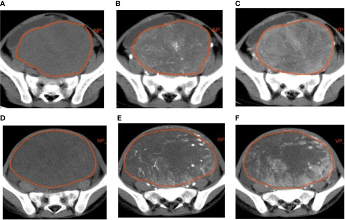
A total of 94 patients with RMS (n = 49) and YSTs (n = 45) were enrolled. Non-enhanced phase (NP), arterial phase (AP), and venous phase (VP) images were retrieved for analysis. The volumes of interest (VOIs) were constructed by segmenting tumor regions on CT images to extract radiomics features. Datasets were randomly divided into two sets including a training set and a test set. In the training set, the least absolute shrinkage and selection operator (LASSO) algorithm was used to screen out the optimal radiomics features that could distinguish RMS from YSTs, and the features were combined with the SVM algorithm to build the classifier model. In the testing set, the areas under the receiver operating characteristic (ROC) curves (AUCs), accuracy, specificity, and sensitivity of the model were calculated to evaluate its diagnostic performance. The clinical factors (including age, sex, tumor site, tumor volume, AFP level) were collected.
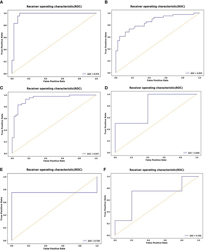
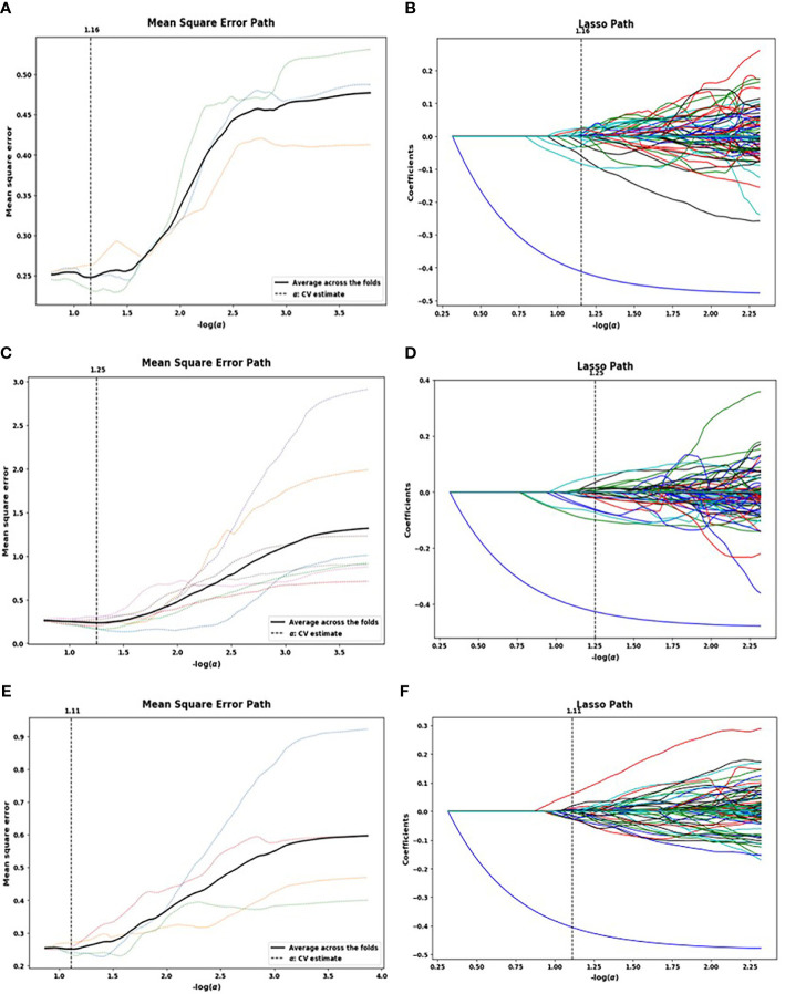
In total, 1,321 features were extracted from the NP, AP, and VP images. The LASSO regression algorithm was used to screen out 23, 26, and 17 related features, respectively. Subsequently, to prevent model overfitting, the 10 features with optimal correlation coefficients were retained. The SVM classifier achieved good diagnostic performance. The AUCs of the NP, AP, and VP radiomics models were 0.937 (95% CI: 0.862, 0.978), 0.973 (95% CI: 0.913, 0.996), and 0.855 (95% CI: 0.762, 0.922) in the training set, respectively, which were confirmed in the test set by AUCs of 0.700 (95% CI: 0.328, 0.940), 0.800 (95% CI: 0.422, 0.979), and 0.750 (95% CI: 0.373, 0.962), respectively. The difference in sex, tumor volume, and AFP level were statistically significant (P < 0.05).
The CT-based radiomics model can be used to effectively distinguish RMS and YST, and combined with clinical features, which can improve diagnostic accuracy and increase the confidence of radiologists in the diagnosis of pelvic solid tumors in children.
本研究旨在探讨CT影像组学特征联合支持向量机(SVM)模型在儿童盆腔横纹肌肉瘤(RMS)与卵黄囊瘤(YST)鉴别诊断中的作用。
共纳入94例RMS患者(n = 49)和YST患者(n = 45)。提取非增强期(NP)、动脉期(AP)和静脉期(VP)图像进行分析。通过在CT图像上分割肿瘤区域构建感兴趣区(VOI)以提取影像组学特征。数据集随机分为训练集和测试集。在训练集中,使用最小绝对收缩和选择算子(LASSO)算法筛选出可区分RMS与YST的最佳影像组学特征,并将这些特征与SVM算法相结合构建分类器模型。在测试集中,计算模型的受试者操作特征(ROC)曲线下面积(AUC)、准确性、特异性和敏感性以评估其诊断性能。收集临床因素(包括年龄、性别、肿瘤部位、肿瘤体积、甲胎蛋白水平)。
共从NP、AP和VP图像中提取了1321个特征。LASSO回归算法分别筛选出23、26和17个相关特征。随后,为防止模型过度拟合,保留了10个相关性最佳的特征。SVM分类器具有良好的诊断性能。训练集中NP、AP和VP影像组学模型的AUC分别为0.937(95%CI:0.862,0.978)、0.973(95%CI:0.913,0.996)和0.855(95%CI:0.762,0.922),测试集中相应的AUC分别为0.700(95%CI:0.328,0.940)、0.800(95%CI:0.422,0.979)和0.750(95%CI:0.373,0.962)。性别、肿瘤体积和甲胎蛋白水平的差异具有统计学意义(P < 0.05)。
基于CT的影像组学模型可有效鉴别RMS和YST,并结合临床特征,可提高诊断准确性,增强放射科医生对儿童盆腔实体肿瘤诊断的信心。