Second Clinical Medicine College, Binzhou Medical University, Yantai, China.
Department of Otorhinolaryngology-Head and Neck Surgery, Yantai Yuhuangding Hospital, Qingdao University, Yantai, China.
Front Endocrinol (Lausanne). 2021 Oct 21;12:741698. doi: 10.3389/fendo.2021.741698. eCollection 2021.
This study aimed to develop a computed tomography (CT)-based radiomics model to predict central lymph node metastases (CLNM) preoperatively in patients with papillary thyroid carcinoma (PTC).
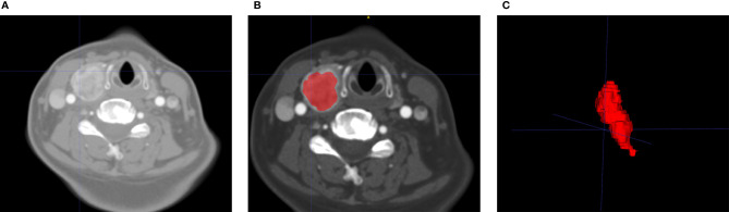
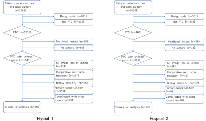
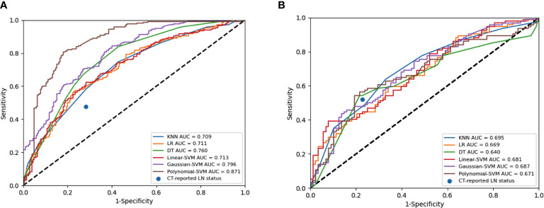
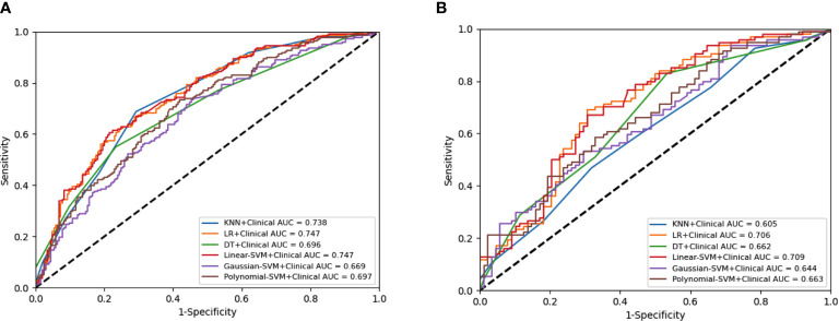
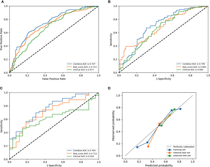
In this retrospective study, 678 patients with PTC were enrolled from Yantai Yuhuangding Hot3spital (n=605) and the Affiliated Hospital of Binzhou Medical University (n=73) within August 2010 to December 2020. The patients were randomly divided into a training set (n=423), an internal test set (n=182), and an external test set (n=73). Radiomics features of each patient were extracted from preoperative plain scan and contrast-enhanced CT images (arterial and venous phases). One-way analysis of variance (ANOVA) and least absolute shrinkage and selection operator algorithm were used for feature selection. The K-nearest neighbor, logistics regression, decision tree, linear-support vector machine (linear-SVM), Gaussian-SVM, and polynomial-SVM algorithms were used to establish radiomics models for CLNM prediction. The clinical risk factors were selected by ANOVA and multivariate logistic regression. Incorporated with clinical risk factors, a combined radiomics model was established for the preoperative prediction of CLNM in patients with PTCs. The performance of the combined radiomics model was evaluated using the receiver operating characteristic (ROC) and calibration curves in the training and test sets. The clinical usefulness was evaluated through decision curve analysis (DCA).
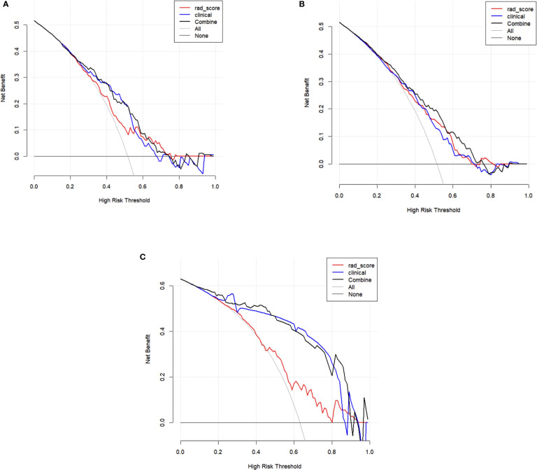
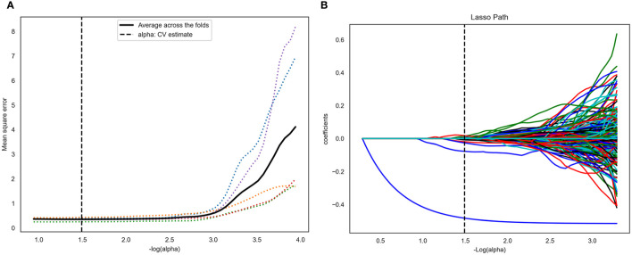
A total of 4227 radiomic features were extracted from the CT images of each patient, and 14 non-zero coefficient features associated with CLNM were selected. Four clinical variables (sex, age, tumor diameter, and CT-reported lymph node status) were significantly associated with CLNM. Linear-SVM led to the best prediction model, which incorporated radiomic features and clinical risk factors. Areas under the ROC curves of 0.747 (95% confidence interval [CI] 0.706-0.782), 0.710 (95% CI 0.634-0.786), and 0.764 (95% CI 0.654-0.875) were obtained in the training, internal, and external test sets, respectively. The linear-SVM algorithm also showed better sensitivity (0.702 [95% CI 0.600-0.790] 0.477 [95% CI 0.409-0.545]) and accuracy (0.670 [95% CI 0.600-0.738] 0.642 [95% CI 0.569-0.712]) than an experienced radiologist in the internal test set in the combined radiomics model. The calibration plot reflected a favorable agreement between the actual and estimated probabilities of CLNM. The DCA indicated the clinical usefulness of the combined radiomics model.
The combined radiomics model is a non-invasive preoperative tool that incorporates radiomic features and clinical risk factors to predict CLNM in patients with PTC.
本研究旨在建立一种基于计算机断层扫描(CT)的放射组学模型,以预测甲状腺乳头状癌(PTC)患者术前中央淋巴结转移(CLNM)。
本回顾性研究纳入了 2010 年 8 月至 2020 年 12 月期间在烟台毓璜顶医院(n=605)和滨州医学院附属医院(n=73)的 678 例 PTC 患者。患者被随机分为训练集(n=423)、内部测试集(n=182)和外部测试集(n=73)。从每位患者的术前平扫和增强 CT 图像(动脉期和静脉期)中提取放射组学特征。使用单因素方差分析(ANOVA)和最小绝对值收缩和选择算子算法进行特征选择。使用 K-最近邻、逻辑回归、决策树、线性支持向量机(linear-SVM)、高斯支持向量机(Gaussian-SVM)和多项式支持向量机(polynomial-SVM)算法建立用于 CLNM 预测的放射组学模型。使用 ANOVA 和多变量逻辑回归选择临床风险因素。将临床风险因素与放射组学模型相结合,建立用于预测 PTC 患者 CLNM 的联合放射组学模型。使用训练集和测试集的受试者工作特征(ROC)曲线和校准曲线评估联合放射组学模型的性能。通过决策曲线分析(DCA)评估临床实用性。
从每位患者的 CT 图像中提取了 4227 个放射组学特征,选择了 14 个与 CLNM 相关的非零系数特征。4 个临床变量(性别、年龄、肿瘤直径和 CT 报告的淋巴结状态)与 CLNM 显著相关。linear-SVM 导致了最佳预测模型,该模型结合了放射组学特征和临床风险因素。在训练、内部和外部测试集中,ROC 曲线下面积分别为 0.747(95%置信区间 [CI] 0.706-0.782)、0.710(95% CI 0.634-0.786)和 0.764(95% CI 0.654-0.875)。linear-SVM 算法在内部测试集中的敏感性(0.702 [95% CI 0.600-0.790] 0.477 [95% CI 0.409-0.545])和准确性(0.670 [95% CI 0.600-0.738] 0.642 [95% CI 0.569-0.712])均优于经验丰富的放射科医生。校准图反映了 CLNM 实际概率和估计概率之间的良好一致性。DCA 表明了联合放射组学模型的临床实用性。
联合放射组学模型是一种非侵入性的术前工具,可结合放射组学特征和临床风险因素预测 PTC 患者的 CLNM。