Son Hee Young, Kim Sanghoon, Mohammad Ramla Talib, Huh Gene, Kim Hyojin, Jeong Woo-Jin, Cha Wonjae
Department of Otorhinolaryngology-Head and Neck Surgery, The Dongnam Institute of Radiological and Medical Sciences (DIRAMS), Busan, Korea.
Department of Otorhinolaryngology-Head and Neck Surgery and Biomedical Research Institute, Pusan National University Hospital, Busan, Korea.
Clin Exp Otorhinolaryngol. 2021 Aug;14(3):338-346. doi: 10.21053/ceo.2020.02180. Epub 2020 Dec 4.
The transcutaneous approach is a good option for office-based vocal fold injection (VFI). However, precise localization requires extensive experience because the needle tip is invisible in small and complex laryngeal spaces. Recently, real-time light-guided VFI (RL-VFI) was proposed as a new technique that allows simultaneous injection under precise needle localization by light guidance. Herein, we aimed to verify the feasibility of RL-VFI in an in vivo canine model and explored its clinical usefulness.
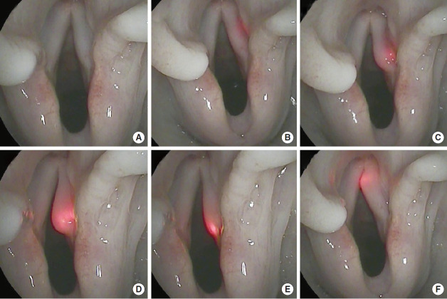
The device for RL-VFI comprised a light source (light-emitting diode modules [10 W] of red color [650 nm]) and injectors (1.5 inches, 23 gauge). An adult male beagle was used for the experiment. After tracheostomy, a rigid laryngoscope was inserted and suspended to expose the larynx. A flexible naso-laryngoscopy system was used to visualize the vocal folds.
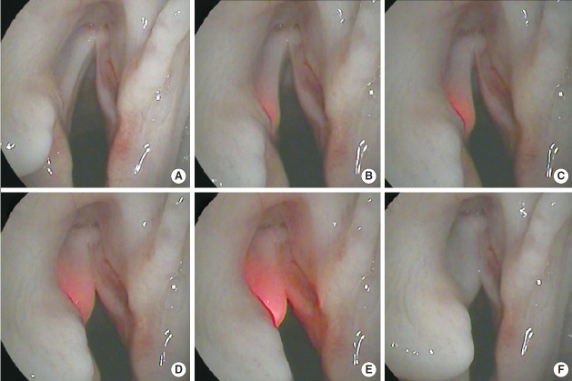
RL-VFI was performed using various transcutaneous approaches, including the cricothyroid, transthyroid, and transhyoid approaches. Light guidance helped identify the path of the needle and prevent inadvertent penetration. The location of the needle tip was accurately indicated by the light. The illuminated needle could be easily placed at the intended points in the vocal fold with real-time visual-motor feedback. Hyaluronic acid could be simultaneously injected lateral to the vocal process under light guidance without manipulation of the device.
RL-VFI was found to be safe and feasible in an in vivo canine model, providing precise localization and visualmotor feedback. The clinical application of RL-VFI is expected to improve the safety and precision of VFI.
经皮途径是门诊声带注射(VFI)的一种良好选择。然而,精确的定位需要丰富的经验,因为在狭小且复杂的喉部空间中针尖是不可见的。最近,实时光导VFI(RL-VFI)作为一种新技术被提出,它能够在光导的精确针尖定位下同时进行注射。在此,我们旨在验证RL-VFI在犬体内模型中的可行性,并探索其临床实用性。
RL-VFI设备包括一个光源(红色[650纳米]发光二极管模块[10瓦])和注射器(1.5英寸,23号)。使用成年雄性比格犬进行实验。气管切开术后,插入并悬吊硬质喉镜以暴露喉部。使用柔性鼻咽喉镜系统观察声带。
使用多种经皮途径进行RL-VFI,包括环甲膜、甲状腺和甲状舌骨途径。光导有助于确定针的路径并防止意外穿透。光精确指示了针尖的位置。在实时视觉运动反馈下,可轻松将发光针放置在声带的预定点。在光导下可同时在声带突外侧注射透明质酸,而无需操作设备。
在犬体内模型中发现RL-VFI安全可行,可提供精确的定位和视觉运动反馈。预计RL-VFI的临床应用将提高VFI的安全性和精确性。