Zaid Mohamed, Elganainy Dalia, Dogra Prashant, Dai Annie, Widmann Lauren, Fernandes Pearl, Wang Zhihui, Pelaez Maria J, Ramirez Javier R, Singhi Aatur D, Dasyam Anil K, Brand Randall E, Park Walter G, Rahmanuddin Syed, Rosenthal Michael H, Wolpin Brian M, Khalaf Natalia, Goel Ajay, Von Hoff Daniel D, Tamm Eric P, Maitra Anirban, Cristini Vittorio, Koay Eugene J
Department of Radiation Oncology, The University of Texas MD Anderson Cancer Center, Houston, TX, United States.
Mathematics in Medicine Program, Houston Methodist Research Institute, Houston, TX, United States.
Front Oncol. 2020 Dec 2;10:596931. doi: 10.3389/fonc.2020.596931. eCollection 2020.
Previously, we characterized subtypes of pancreatic ductal adenocarcinoma (PDAC) on computed-tomography (CT) scans, whereby conspicuous (high delta) PDAC tumors are more likely to have aggressive biology and poorer clinical outcomes compared to inconspicuous (low delta) tumors. Here, we hypothesized that these imaging-based subtypes would exhibit different growth-rates and distinctive metabolic effects in the period prior to PDAC diagnosis.
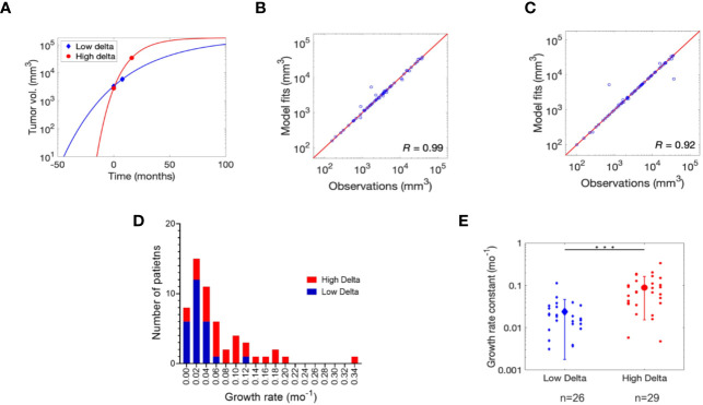
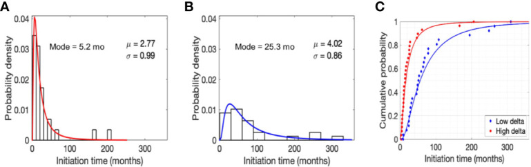
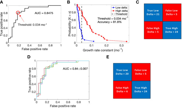
Retrospectively, we evaluated 55 patients who developed PDAC as a second primary cancer and underwent serial pre-diagnostic (T0) and diagnostic (T1) CT-scans. We scored the PDAC tumors into high and low delta on T1 and, serially, obtained the biaxial measurements of the pancreatic lesions (T0-T1). We used the Gompertz-function to model the growth-kinetics and estimate the tumor growth-rate constant (α) which was used for tumor binary classification, followed by cross-validation of the classifier accuracy. We used maximum-likelihood estimation to estimate initiation-time from a single cell (10 mm) to a 10 mm tumor mass. Finally, we serially quantified the subcutaneous-abdominal-fat (SAF), visceral-abdominal-fat (VAF), and muscles volumes (cm) on CT-scans, and recorded the change in blood glucose (BG) levels. T-test, likelihood-ratio, Cox proportional-hazards, and Kaplan-Meier were used for statistical analysis and p-value <0.05 was considered significant.
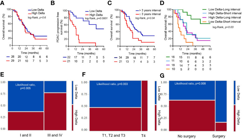
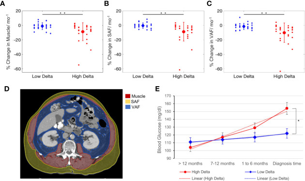
Compared to high delta tumors, low delta tumors had significantly slower average growth-rate constants (0.024 month vs. 0.088 month, p<0.0001) and longer average initiation-times (14 years vs. 5 years, p<0.0001). α demonstrated high accuracy (area under the curve (AUC)=0.85) in classifying the tumors into high and low delta, with an optimal cut-off of 0.034 month. Leave-one-out-cross-validation showed 80% accuracy in predicting the delta-class (AUC=0.84). High delta tumors exhibited accelerated SAF, VAF, and muscle wasting (p <0.001), and BG disturbance (p<0.01) compared to low delta tumors. Patients with low delta tumors had better PDAC-specific progression-free survival (log-rank, p<0.0001), earlier stage tumors (p=0.005), and higher likelihood to receive resection after PDAC diagnosis (p=0.008), compared to those with high delta tumors.
Imaging-based subtypes of PDAC exhibit distinct growth, metabolic, and clinical profiles during the pre-diagnostic period. Our results suggest that heterogeneous disease biology may be an important consideration in early detection strategies for PDAC.
此前,我们在计算机断层扫描(CT)上对胰腺导管腺癌(PDAC)的亚型进行了特征描述,与不明显(低delta)的肿瘤相比,明显(高delta)的PDAC肿瘤更有可能具有侵袭性生物学行为和较差的临床结果。在此,我们假设这些基于影像学的亚型在PDAC诊断前的时期会表现出不同的生长速度和独特的代谢效应。
我们回顾性评估了55例发生PDAC作为第二原发性癌症并接受系列诊断前(T0)和诊断(T1)CT扫描的患者。我们在T1时将PDAC肿瘤分为高delta和低delta,并连续获取胰腺病变的双轴测量值(T0 - T1)。我们使用Gompertz函数对生长动力学进行建模并估计肿瘤生长速率常数(α),该常数用于肿瘤二元分类,随后对分类器准确性进行交叉验证。我们使用最大似然估计来估计从单个细胞(10毫米)到10毫米肿瘤块的起始时间。最后,我们在CT扫描上连续量化皮下腹部脂肪(SAF)、内脏腹部脂肪(VAF)和肌肉体积(厘米),并记录血糖(BG)水平的变化。使用t检验、似然比、Cox比例风险和Kaplan - Meier进行统计分析,p值<0.05被认为具有统计学意义。
与高delta肿瘤相比,低delta肿瘤的平均生长速率常数明显较慢(0.024个月对0.088个月,p<0.0001),平均起始时间更长(14年对5年,p<0.0001)。α在将肿瘤分为高delta和低delta方面表现出较高的准确性(曲线下面积(AUC)=0.85),最佳截断值为0.034个月。留一法交叉验证显示在预测delta类别方面的准确率为80%(AUC = 0.84)。与低delta肿瘤相比,高delta肿瘤表现出皮下腹部脂肪、内脏腹部脂肪和肌肉加速消耗(p <0.001)以及血糖紊乱(p<0.01)。与高delta肿瘤患者相比,低delta肿瘤患者的PDAC特异性无进展生存期更好(对数秩检验,p<0.0001),肿瘤分期更早(p = 0.005),并且在PDAC诊断后接受切除的可能性更高(p = 0.008)。
基于影像学的PDAC亚型在诊断前期表现出不同的生长、代谢和临床特征。我们的结果表明,疾病生物学异质性可能是PDAC早期检测策略中的一个重要考虑因素。