Institut NeuroMyoGène - CNRS UMR 5310 - INSERM U1217 de Lyon- UCBL Lyon 1, Faculté de Médecine et de Pharmacie, Lyon, France.
Interdisciplinary Institute for Neuroscience, UMR CNRS 5297 - University of Bordeaux, Bordeaux, France.
Elife. 2020 Dec 21;9:e63205. doi: 10.7554/eLife.63205.
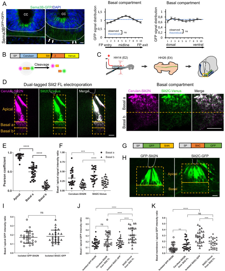
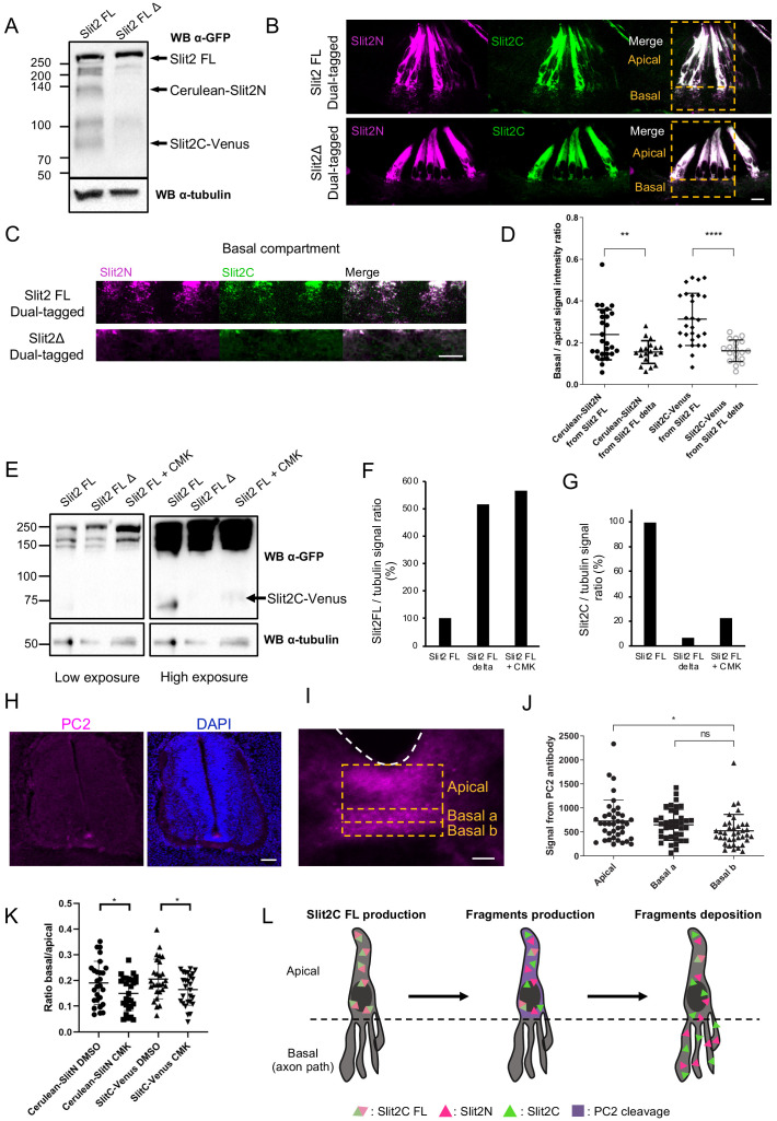
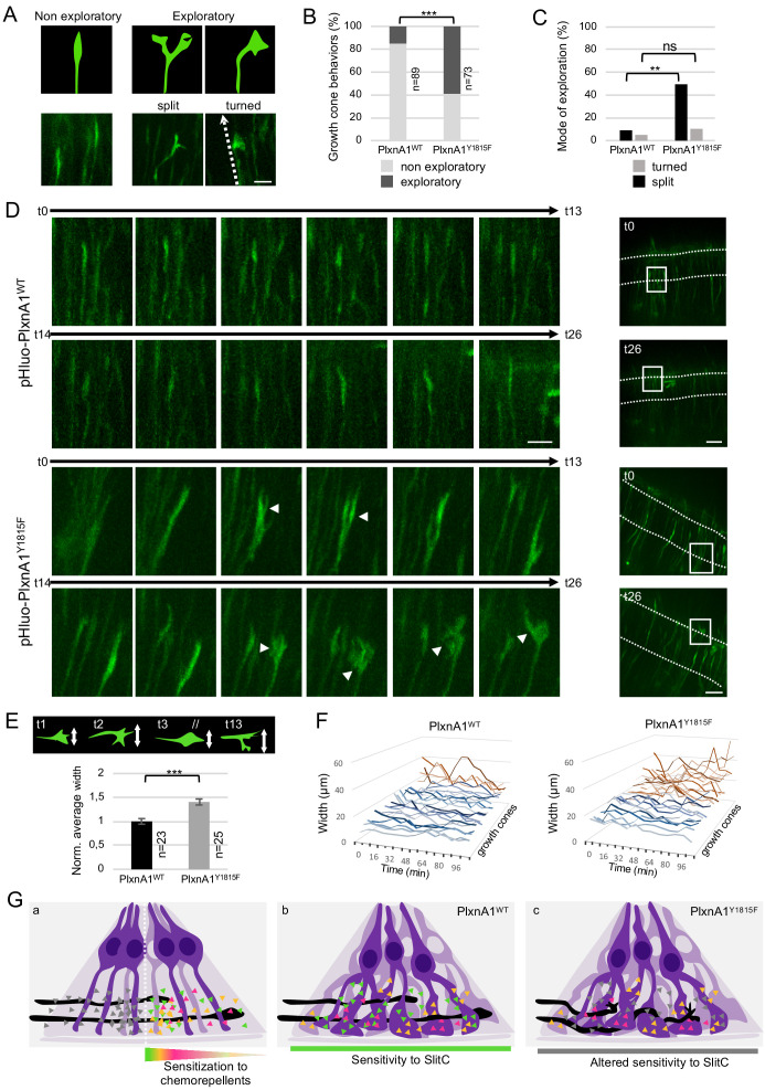
Spinal commissural axon navigation across the midline in the floor plate requires repulsive forces from local Slit repellents. The long-held view is that Slits push growth cones forward and prevent them from turning back once they became sensitized to these cues after midline crossing. We analyzed with fluorescent reporters Slits distribution and FP glia morphology. We observed clusters of Slit-N and Slit-C fragments decorating a complex architecture of glial basal process ramifications. We found that PC2 proprotein convertase activity contributes to this pattern of ligands. Next, we studied Slit-C acting via PlexinA1 receptor shared with another FP repellent, the Semaphorin3B, through generation of a mouse model baring PlexinA1 mutation abrogating SlitC but not Sema3B responsiveness, manipulations in the chicken embryo, and ex vivo live imaging. This revealed a guidance mechanism by which SlitC constantly limits growth cone exploration, imposing ordered and forward-directed progression through aligned corridors formed by FP basal ramifications.
脊髓连合轴突在中线穿过基板需要来自局部 Slit 排斥物的排斥力。长期以来的观点是,Slit 推动生长锥向前移动,并防止它们在中线穿过后对这些线索变得敏感时回头。我们用荧光报告基因分析了 Slit 的分布和 FP 神经胶质形态。我们观察到 Slit-N 和 Slit-C 片段簇装饰着神经胶质基底突起分支的复杂结构。我们发现 PC2 蛋白原转化酶活性有助于这种配体模式。接下来,我们通过生成一种带有 PlexinA1 突变的小鼠模型,该突变消除了 SlitC 但不影响 Sema3B 反应性,在鸡胚中进行操作,并进行离体活体成像,研究了 Slit-C 通过与另一种 FP 排斥物 Semaphorin3B 共享的 PlexinA1 受体发挥作用。这揭示了一种指导机制,通过该机制,SlitC 不断限制生长锥的探索,通过 FP 基底突起分支形成的对齐通道施加有序和向前的定向推进。