Dyer Kathleen I C, Sanfilippo Paul G, White Scott W, Guggenheim Jeremy A, Hammond Chris J, Newnham John P, Mackey David A, Yazar Seyhan
Centre for Ophthalmology and Visual Science, Lions Eye Institute, University of Western Australia, Perth, Western Australia, Australia.
School of Medicine, Faculty of Health and Medical Sciences, University of Western Australia, Perth, Western Australia, Australia.
Invest Ophthalmol Vis Sci. 2020 Dec 1;61(14):26. doi: 10.1167/iovs.61.14.26.
To evaluate the contribution of genetic and early life environmental factors, as reflected by fetal anthropometric growth trajectories, toward the development of myopia during childhood and adolescence.
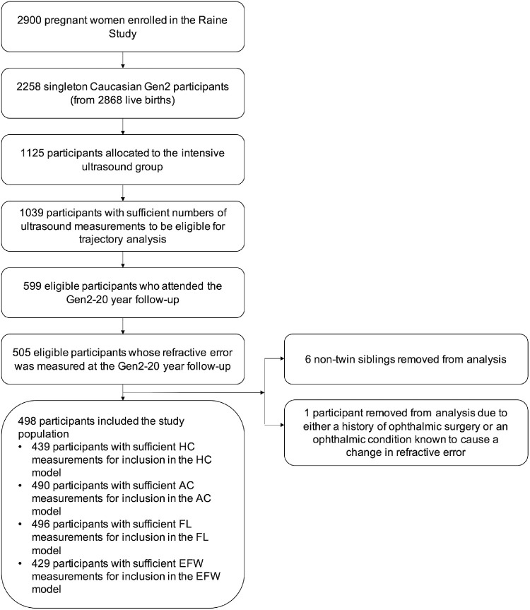
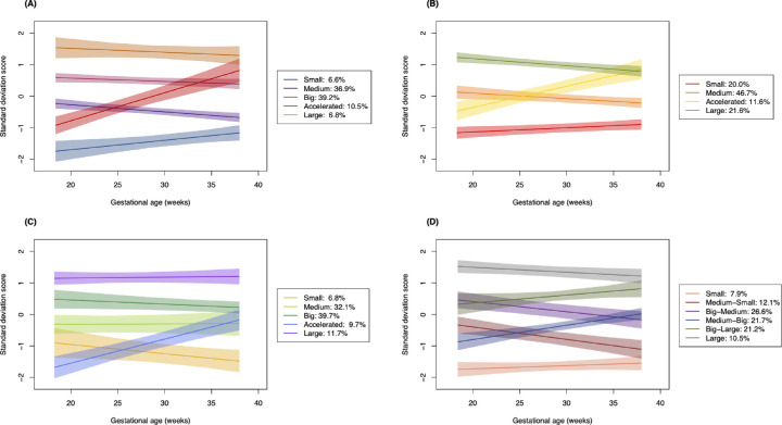
This analysis included 498 singleton Caucasian participants from the Raine Study, a pregnancy cohort study based in Western Australia. Serial fetal biometric measurements of these participants were collected via ultrasound scans performed at 18, 24, 28, 34, and 38 weeks' gestation. At a 20-year follow-up, the participants underwent a comprehensive ophthalmic examination, including cycloplegic autorefraction and ocular biometry measurements. Using a group-based trajectory modeling approach, we identified groups of participants with similar growth trajectories based on measurements of fetal head circumference (HC), abdominal circumference, femur length (FL), and estimated fetal weight (EFW). Differences between trajectory groups with respect to prevalence of myopia, axial length (AL), and corneal radius of curvature measured at the 20-year follow-up were evaluated via logistic regression and analysis of variance.
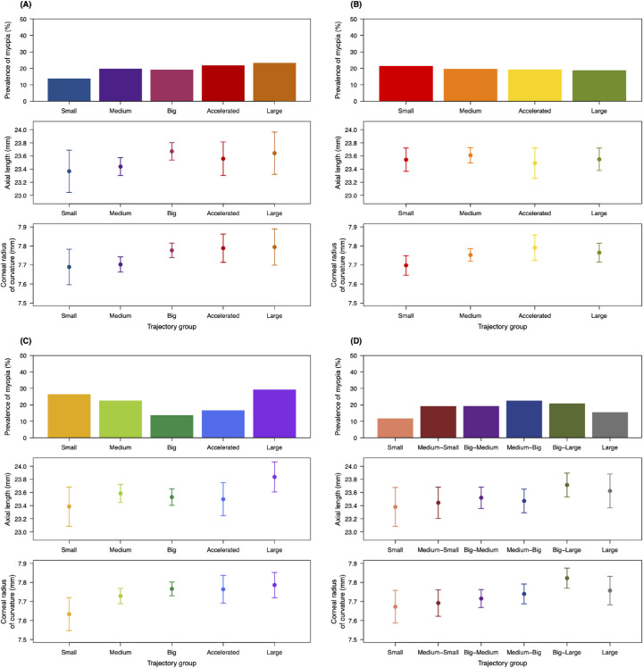
Prevalence of myopia was highest among participants with consistently short or consistently long FLs (P = 0.04). There was also a trend toward increased prevalence with larger HC in late gestation, although not at a statistically significant level. Trajectory groups reflecting faster HC, FL, or EFW growth correlated with significantly flatter corneas (P = 0.03, P = 0.04, and P = 0.01, respectively) and a general, but not statistically significant, increase in AL.
Environmental or genetic factors influencing intrauterine skeletal growth may concurrently affect ocular development, with effects persisting into adulthood.
评估胎儿人体测量生长轨迹所反映的遗传和早期生活环境因素对儿童及青少年近视发展的影响。
本分析纳入了来自西澳大利亚一项妊娠队列研究——雷恩研究的498名单胎白种人参与者。通过在妊娠18、24、28、34和38周时进行的超声扫描收集这些参与者的系列胎儿生物测量数据。在20年随访时,参与者接受了全面的眼科检查,包括睫状肌麻痹验光和眼生物测量。使用基于群体的轨迹建模方法,我们根据胎儿头围(HC)、腹围、股骨长度(FL)和估计胎儿体重(EFW)的测量结果,确定了具有相似生长轨迹的参与者群体。通过逻辑回归和方差分析评估了轨迹组在20年随访时近视患病率、眼轴长度(AL)和角膜曲率半径方面的差异。
FL持续短或持续长的参与者中近视患病率最高(P = 0.04)。妊娠晚期HC较大时,近视患病率也有增加趋势,尽管未达到统计学显著水平。反映HC、FL或EFW生长较快的轨迹组与角膜明显更扁平相关(分别为P = 0.03、P = 0.04和P = 0.01),AL总体上有增加但无统计学显著意义。
影响子宫内骨骼生长的环境或遗传因素可能同时影响眼部发育,且这种影响会持续到成年期。