• 文献检索
  • 文档翻译
  • 深度研究
  • 学术资讯
  • Suppr Zotero 插件Zotero 插件
  • 邀请有礼
  • 套餐&价格
  • 历史记录
应用&插件
Suppr Zotero 插件Zotero 插件浏览器插件Mac 客户端Windows 客户端微信小程序
定价
高级版会员购买积分包购买API积分包
服务
文献检索文档翻译深度研究API 文档MCP 服务
关于我们
关于 Suppr公司介绍联系我们用户协议隐私条款
关注我们

Suppr 超能文献

核心技术专利:CN118964589B侵权必究
粤ICP备2023148730 号-1Suppr @ 2026

文献检索

告别复杂PubMed语法,用中文像聊天一样搜索,搜遍4000万医学文献。AI智能推荐,让科研检索更轻松。

立即免费搜索

文件翻译

保留排版,准确专业,支持PDF/Word/PPT等文件格式,支持 12+语言互译。

免费翻译文档

深度研究

AI帮你快速写综述,25分钟生成高质量综述,智能提取关键信息,辅助科研写作。

立即免费体验

影像学 - 超声生物显微镜和眼前节光学相干断层扫描 - 在眼前节病变的诊断中的应用。

Imagery - ultrasound biomicroscopy and anterior segment Optical Coherence Tomography - in the diagnosis of anterior segment pathology.

机构信息

"Dr. Carol Davila" Central Military Emergency Hospital, Bucharest, Romania.

"Carol Davila" University of Medicine and Pharmacy, Bucharest, Romania.

出版信息

Rom J Ophthalmol. 2020 Jul-Sep;64(3):292-298.

PMID:33367163
原文链接:https://pmc.ncbi.nlm.nih.gov/articles/PMC7739559/
Abstract

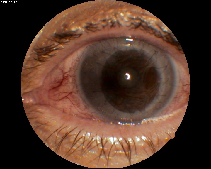
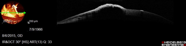
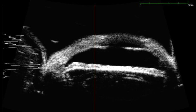
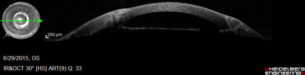
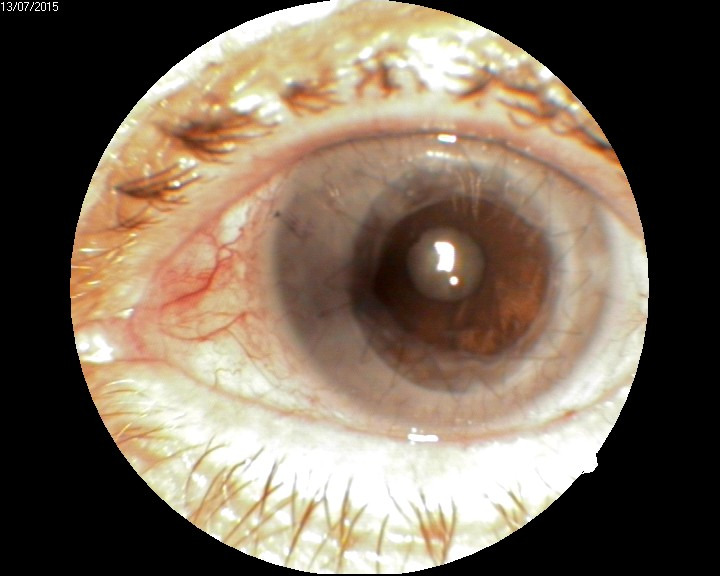
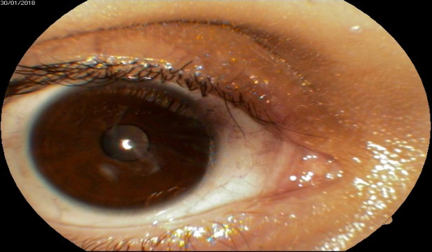
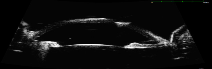
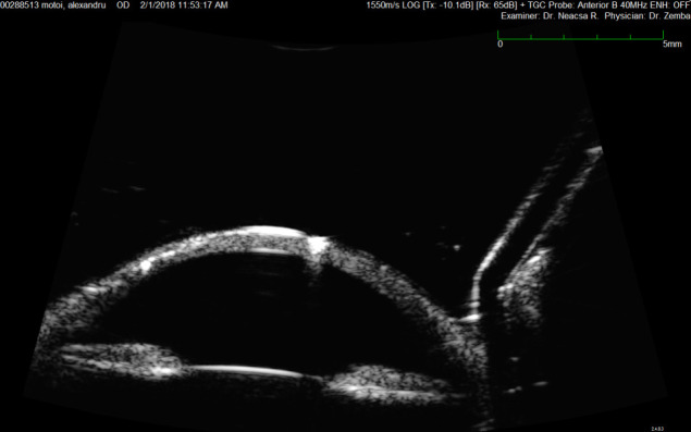
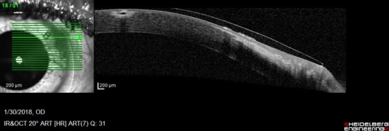
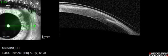
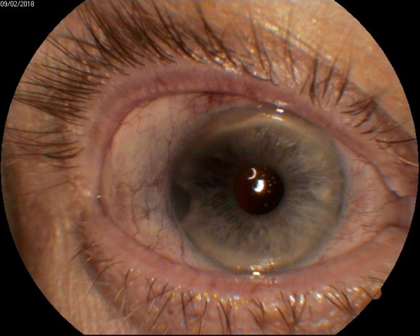
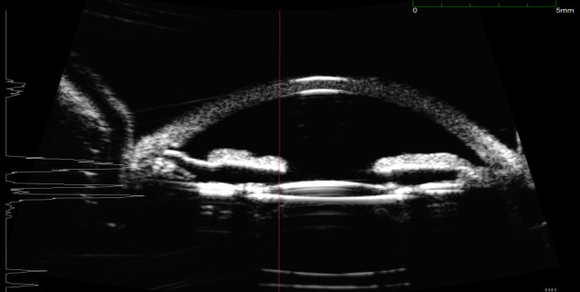
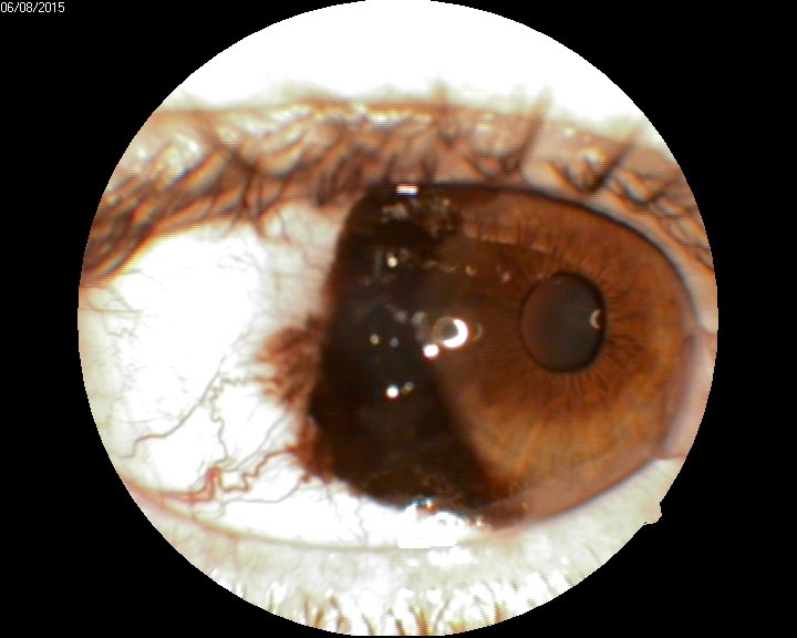
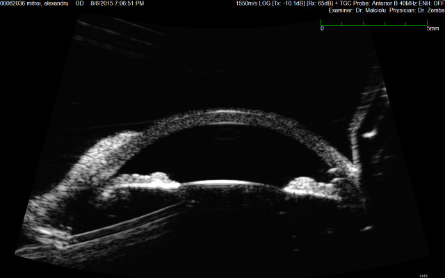
The aim of this paper was to show the usefulness of imagery in better documenting the pathology of the anterior segment. The article comprises clinical cases, insisting on how imagery was essential in establishing the diagnosis or the therapeutic plan. Lack of imagery would have made establishing a proper diagnosis much more difficult. Although clinical examination is simple and offers a fairly good amount of information, some particular cases of anterior segment pathology need additional investigations, every method having its indications and limits.

摘要

本文旨在展示影像学在更好地记录眼前节病理学方面的作用。文章包含临床病例,强调了影像学在确定诊断或治疗计划方面的重要性。如果没有影像学,正确诊断的难度将会大大增加。虽然临床检查简单,提供了相当多的信息,但有些特定的眼前节病理学病例需要额外的检查,每种方法都有其适应证和局限性。

https://cdn.ncbi.nlm.nih.gov/pmc/blobs/5778/7739559/c886e1ddd2ce/RomJOphthalmol-64-292-g0014.jpg
https://cdn.ncbi.nlm.nih.gov/pmc/blobs/5778/7739559/0c69f88e2f04/RomJOphthalmol-64-292-g001.jpg
https://cdn.ncbi.nlm.nih.gov/pmc/blobs/5778/7739559/7295c8c18d81/RomJOphthalmol-64-292-g002.jpg
https://cdn.ncbi.nlm.nih.gov/pmc/blobs/5778/7739559/eb758fceae4a/RomJOphthalmol-64-292-g003.jpg
https://cdn.ncbi.nlm.nih.gov/pmc/blobs/5778/7739559/3c114fffd46b/RomJOphthalmol-64-292-g004.jpg
https://cdn.ncbi.nlm.nih.gov/pmc/blobs/5778/7739559/e6c5cc579e80/RomJOphthalmol-64-292-g005.jpg
https://cdn.ncbi.nlm.nih.gov/pmc/blobs/5778/7739559/9570a5867aa6/RomJOphthalmol-64-292-g006.jpg
https://cdn.ncbi.nlm.nih.gov/pmc/blobs/5778/7739559/a57f2a1f0218/RomJOphthalmol-64-292-g007.jpg
https://cdn.ncbi.nlm.nih.gov/pmc/blobs/5778/7739559/cf43f63feb94/RomJOphthalmol-64-292-g008.jpg
https://cdn.ncbi.nlm.nih.gov/pmc/blobs/5778/7739559/3ee7e2165cbd/RomJOphthalmol-64-292-g009.jpg
https://cdn.ncbi.nlm.nih.gov/pmc/blobs/5778/7739559/b4d417bf49e0/RomJOphthalmol-64-292-g0010.jpg
https://cdn.ncbi.nlm.nih.gov/pmc/blobs/5778/7739559/e88be0b760a8/RomJOphthalmol-64-292-g0011.jpg
https://cdn.ncbi.nlm.nih.gov/pmc/blobs/5778/7739559/8e15f1cb9c86/RomJOphthalmol-64-292-g0012.jpg
https://cdn.ncbi.nlm.nih.gov/pmc/blobs/5778/7739559/954441ca0775/RomJOphthalmol-64-292-g0013.jpg
https://cdn.ncbi.nlm.nih.gov/pmc/blobs/5778/7739559/c886e1ddd2ce/RomJOphthalmol-64-292-g0014.jpg

相似文献

1
Imagery - ultrasound biomicroscopy and anterior segment Optical Coherence Tomography - in the diagnosis of anterior segment pathology.影像学 - 超声生物显微镜和眼前节光学相干断层扫描 - 在眼前节病变的诊断中的应用。
Rom J Ophthalmol. 2020 Jul-Sep;64(3):292-298.
2
Anterior segment imaging: optical coherence tomography versus ultrasound biomicroscopy.眼前节成像:光学相干断层扫描与超声生物显微镜检查的对比
Ophthalmic Surg Lasers Imaging. 2008 Nov-Dec;39(6):476-84. doi: 10.3928/15428877-20081101-02.
3
Anterior segment imaging in glaucoma: An updated review.青光眼的眼前段成像:最新综述。
Indian J Ophthalmol. 2015 Aug;63(8):630-40. doi: 10.4103/0301-4738.169787.
4
Imaging the Canaliculops With Ultrasound Biomicroscopy and Anterior Segment Ocular Coherence Tomography.用超声生物显微镜和眼前节光学相干断层扫描对泪小管异常进行成像。
Ophthalmic Plast Reconstr Surg. 2017 Nov/Dec;33(6):e143-e144. doi: 10.1097/IOP.0000000000000878.
5
Anterior segment imaging: ultrasound biomicroscopy and anterior segment optical coherence tomography.眼前节成像:超声生物显微镜检查和眼前节光学相干断层扫描
Curr Opin Ophthalmol. 2008 Mar;19(2):115-21. doi: 10.1097/ICU.0b013e3282f40bba.
6
Advances in anterior segment imaging.眼前节成像的进展
Curr Opin Ophthalmol. 2007 Feb;18(1):32-8. doi: 10.1097/ICU.0b013e328011550d.
7
Anterior segment optical coherence tomography and ultrasound biomicroscopy in the imaging of anterior segment tumors.眼前节光学相干断层扫描和超声生物显微镜在前节肿瘤成像中的应用
Am J Ophthalmol. 2009 Feb;147(2):214-219.e2. doi: 10.1016/j.ajo.2008.08.023. Epub 2008 Oct 18.
8
Comparison of immersion ultrasonography, ultrasound biomicroscopy and anterior segment optical coherence tomography in the evaluation of traumatic phacoceles.比较浸水超声、超声生物显微镜和眼前节光学相干断层扫描在创伤性白内障中的评估。
Indian J Ophthalmol. 2012 Jan-Feb;60(1):63-5. doi: 10.4103/0301-4738.91352.
9
Correlation of anterior segment optical coherence tomography and ultrasound biomicroscopy in congenital corneal opacity.先天性角膜混浊的眼前节光学相干断层扫描与超声生物显微镜的相关性。
J AAPOS. 2024 Apr;28(2):103863. doi: 10.1016/j.jaapos.2024.103863. Epub 2024 Mar 6.
10
Anterior segment imaging using optical coherence tomography and ultrasound biomicroscopy in secondary pigmentary glaucoma associated with in-the-bag intraocular lens.使用光学相干断层扫描和超声生物显微镜对与囊内人工晶状体相关的继发性色素性青光眼进行眼前节成像。
J Cataract Refract Surg. 2006 Nov;32(11):1866-9. doi: 10.1016/j.jcrs.2006.08.018.

引用本文的文献

1
Application of CT Scan in Diagnosis of Iliac-Femoral Vein Thrombosis after Hip Replacement.CT 扫描在髋关节置换术后髂股静脉血栓诊断中的应用。
Scanning. 2022 Jun 20;2022:8428963. doi: 10.1155/2022/8428963. eCollection 2022.

本文引用的文献

1
Retrocorneal membranes after penetrating keratoplasty.穿透性角膜移植术后的角膜后膜
Rom J Ophthalmol. 2015 Oct-Dec;59(4):230-234.
2
Ultra high-resolution anterior segment optical coherence tomography in the evaluation of anterior corneal dystrophies and degenerations.超高分辨率眼前节光学相干断层扫描在评价前部角膜营养不良和变性中的应用。
Ophthalmology. 2011 Jul;118(7):1291-6. doi: 10.1016/j.ophtha.2010.12.015. Epub 2011 Mar 21.
3
Intrastromal corneal ring segment SK implantation for moderate to severe keratoconus.基质内角膜环段 SK 植入术治疗中重度圆锥角膜。
J Cataract Refract Surg. 2010 Jan;36(1):110-3. doi: 10.1016/j.jcrs.2009.07.040.
4
Clinical and research applications of anterior segment optical coherence tomography - a review.眼前节光学相干断层扫描的临床与研究应用——综述
Clin Exp Ophthalmol. 2009 Jan;37(1):81-9. doi: 10.1111/j.1442-9071.2008.01823.x. Epub 2008 Nov 5.
5
Clinical features distinguishing angle closure from pseudoplateau versus plateau iris.区分闭角型青光眼与假性房角劈裂和房角高原征的临床特征。
Br J Ophthalmol. 2008 Mar;92(3):340-4. doi: 10.1136/bjo.2007.114876. Epub 2008 Jan 22.
6
Anterior chamber width measurement by high-speed optical coherence tomography.通过高速光学相干断层扫描测量前房宽度
Ophthalmology. 2005 Feb;112(2):238-44. doi: 10.1016/j.ophtha.2004.09.019.
7
Ultrasound biomicroscopy in management of malignant iris melanoma.超声生物显微镜在恶性虹膜黑色素瘤治疗中的应用
Arch Ophthalmol. 2003 May;121(5):725-7. doi: 10.1001/archopht.121.5.725.
8
Ultrasound biomicroscopy as a tool for detecting and localizing occult foreign bodies after ocular trauma.超声生物显微镜作为检测和定位眼外伤后隐匿性异物的工具。
Ophthalmology. 1999 Feb;106(2):301-5. doi: 10.1016/S0161-6420(99)90056-8.
9
Ultrasound biomicroscopic quantitative analysis of light-dark changes associated with pupillary block.超声生物显微镜对与瞳孔阻滞相关的明暗变化的定量分析
Am J Ophthalmol. 1999 Jan;127(1):43-7. doi: 10.1016/s0002-9394(98)00283-9.
10
Ultrasound biomicroscopy in anterior ocular trauma.眼前部外伤的超声生物显微镜检查
Ophthalmic Surg Lasers. 1997 Mar;28(3):201-7.