Alhasyimi Ananto Ali, Suparwitri Sri, Christnawati Christnawati
Department of Orthodontics, Faculty of Dentistry, Universitas Gadjah Mada, Yogyakarta, Indonesia.
Eur J Dent. 2021 Jul;15(3):412-419. doi: 10.1055/s-0040-1721234. Epub 2020 Dec 26.
This study aimed to determine the effect of carbonate apatite (CHA) hydrogel-aPRF on osteoblastogenesis during relapse in rabbits.
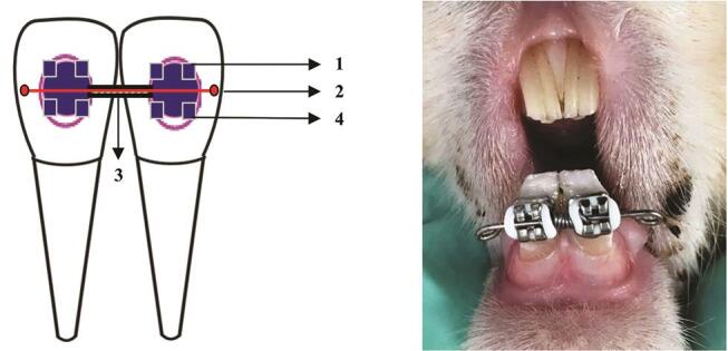
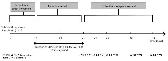
Forty-five rabbits were divided into three groups ( = 15): the control, CHA, and CHA-autologous platelet-rich fibrin (aPRF) groups. An open-coil spring was compressed between brackets to distalize the lower incisors of the rabbits by delivering a force of 50 cN for 1 week. The new position of the teeth was retained for 14 days, and CHA hydrogel-aPRF was injected every 7 days. The appliances were then debonded to allow relapse. On days 0, 3, 7, 14, and 21 after debonding, transforming growth factor (TGF)-β1 and bone morphogenetic protein (BMP)-2 expression was examined using immunohistochemistry staining and Runx-2 levels were analyzed by enzyme-linked immunosorbent assay. The data collected were analyzed using analysis of variance and a post hoc Tukey's test ( < 0.05).
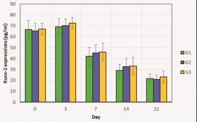
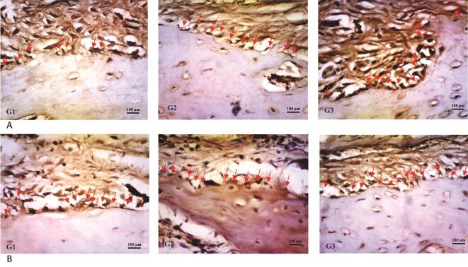
Histomorphometric analysis revealed that TGF-β1 expression in the CHA-aPRF group is statistically higher than that in other groups on days 0, 3, and 7 after debonding ( < 0.05). BMP-2 expression in the CHA-aPRF group was also statistically higher than that in the other groups on days 3, 14, and 21 after debonding ( < 0.05). ELISA showed that Runx-2 levels are slightly higher in the CHA-aPRF group than in the other groups ( > 0.05).
Although injection of CHA-aPRF aids in osteoblastogenesis associated with enhancing TGF-β1 and BMP-2 expressions, it does not significantly upregulate Runx-2 levels.
本研究旨在确定碳酸磷灰石(CHA)水凝胶 - 自体富血小板纤维蛋白(aPRF)对兔复发过程中成骨细胞生成的影响。
45只兔子分为三组(每组n = 15):对照组、CHA组和CHA - 自体富血小板纤维蛋白(aPRF)组。在托槽之间压缩一个开圈弹簧,通过施加50 cN的力使兔子的下切牙远中移动1周。牙齿的新位置保持14天,每7天注射CHA水凝胶 - aPRF。然后拆除矫治器以允许复发。在拆除矫治器后的第0、3、7、14和21天,使用免疫组织化学染色检测转化生长因子(TGF) - β1和骨形态发生蛋白(BMP) - 2的表达,并通过酶联免疫吸附测定分析Runx - 2水平。收集的数据使用方差分析和事后Tukey检验进行分析(P < 0.05)。
组织形态计量学分析显示,在拆除矫治器后的第0、3和7天,CHA - aPRF组中TGF - β1的表达在统计学上高于其他组(P < 0.05)。在拆除矫治器后的第3、14和21天,CHA - aPRF组中BMP - 2的表达在统计学上也高于其他组(P < 0.05)。酶联免疫吸附测定显示,CHA - aPRF组中Runx - 2水平略高于其他组(P > 0.05)。
尽管注射CHA - aPRF有助于与增强TGF - β1和BMP - 2表达相关的成骨细胞生成,但它并未显著上调Runx - 2水平。