• 文献检索
  • 文档翻译
  • 深度研究
  • 学术资讯
  • Suppr Zotero 插件Zotero 插件
  • 邀请有礼
  • 套餐&价格
  • 历史记录
应用&插件
Suppr Zotero 插件Zotero 插件浏览器插件Mac 客户端Windows 客户端微信小程序
定价
高级版会员购买积分包购买API积分包
服务
文献检索文档翻译深度研究API 文档MCP 服务
关于我们
关于 Suppr公司介绍联系我们用户协议隐私条款
关注我们

Suppr 超能文献

核心技术专利:CN118964589B侵权必究
粤ICP备2023148730 号-1Suppr @ 2026

文献检索

告别复杂PubMed语法,用中文像聊天一样搜索,搜遍4000万医学文献。AI智能推荐,让科研检索更轻松。

立即免费搜索

文件翻译

保留排版,准确专业,支持PDF/Word/PPT等文件格式,支持 12+语言互译。

免费翻译文档

深度研究

AI帮你快速写综述,25分钟生成高质量综述,智能提取关键信息,辅助科研写作。

立即免费体验

动态[C]PE2I PET-MR脑成像中零回波时间、单图谱和多图谱衰减校正的准确性和精确性

Accuracy and precision of zero-echo-time, single- and multi-atlas attenuation correction for dynamic [C]PE2I PET-MR brain imaging.

作者信息

Sousa João M, Appel Lieuwe, Merida Inés, Heckemann Rolf A, Costes Nicolas, Engström Mathias, Papadimitriou Stergios, Nyholm Dag, Ahlström Håkan, Hammers Alexander, Lubberink Mark

机构信息

Department of Surgical Sciences, Uppsala University, Uppsala, Sweden.

Medical Imaging Centre, Uppsala University Hospital, Uppsala, Sweden.

出版信息

EJNMMI Phys. 2020 Dec 28;7(1):77. doi: 10.1186/s40658-020-00347-2.

DOI:10.1186/s40658-020-00347-2
PMID:33369700
原文链接:https://pmc.ncbi.nlm.nih.gov/articles/PMC7769756/
Abstract

BACKGROUND

A valid photon attenuation correction (AC) method is instrumental for obtaining quantitatively correct PET images. Integrated PET/MR systems provide no direct information on attenuation, and novel methods for MR-based AC (MRAC) are still under investigation. Evaluations of various AC methods have mainly focused on static brain PET acquisitions. In this study, we determined the validity of three MRAC methods in a dynamic PET/MR study of the brain.

METHODS

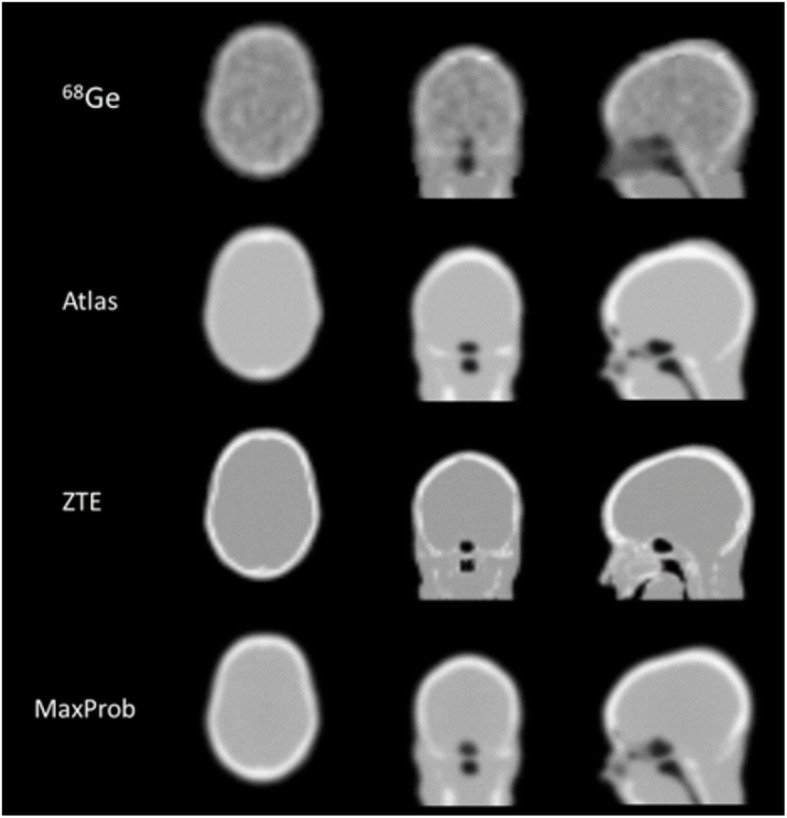
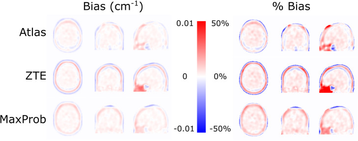
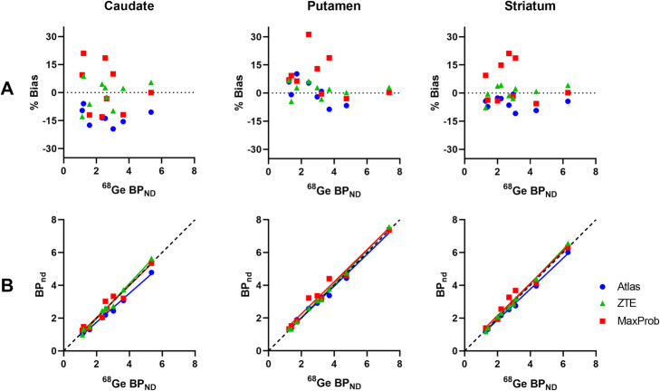
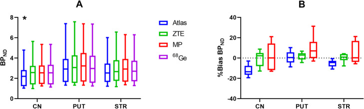
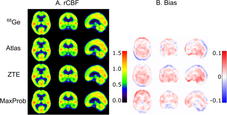
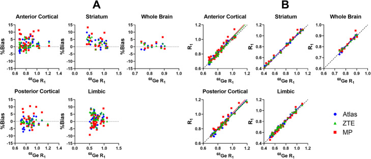
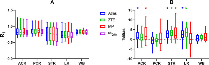
Nine participants underwent dynamic brain PET/MR scanning using the dopamine transporter radioligand [C]PE2I. Three MRAC methods were evaluated: single-atlas (Atlas), multi-atlas (MaxProb) and zero-echo-time (ZTE). The Ge-transmission data from a previous stand-alone PET scan was used as reference method. Parametric relative delivery (R) images and binding potential (BP) maps were generated using cerebellar grey matter as reference region. Evaluation was based on bias in MRAC maps, accuracy and precision of [C]PE2I BP and R estimates, and [C]PE2I time-activity curves. BP was examined for striatal regions and R in clusters of regions across the brain.

RESULTS

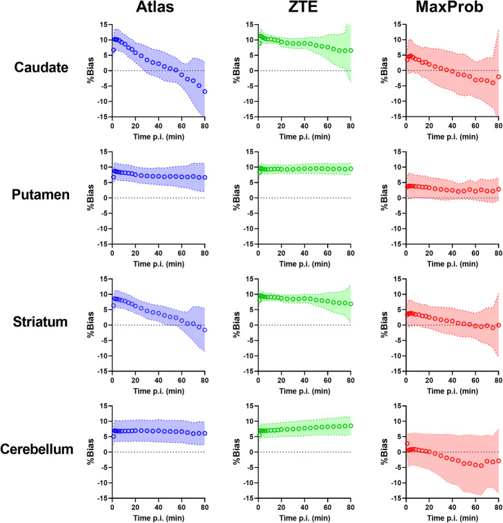
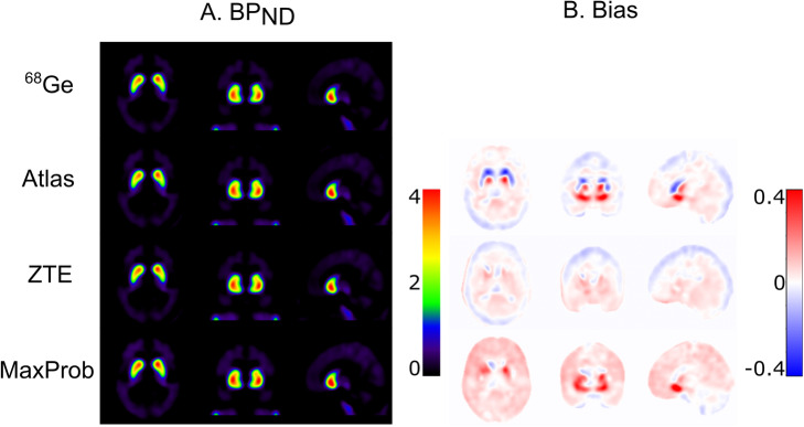
For BP, ZTE-MRAC showed the highest accuracy (bias < 2%) in striatal regions. Atlas-MRAC exhibited a significant bias in caudate nucleus (- 12%) while MaxProb-MRAC revealed a substantial, non-significant bias in the putamen (9%). R estimates had a marginal bias for all MRAC methods (- 1.0-3.2%). MaxProb-MRAC showed the largest intersubject variability for both R and BP. Standardized uptake values (SUV) of striatal regions displayed the strongest average bias for ZTE-MRAC (~ 10%), although constant over time and with the smallest intersubject variability. Atlas-MRAC had highest variation in bias over time (+10 to - 10%), followed by MaxProb-MRAC (+5 to - 5%), but MaxProb showed the lowest mean bias. For the cerebellum, MaxProb-MRAC showed the highest variability while bias was constant over time for Atlas- and ZTE-MRAC.

CONCLUSIONS

Both Maxprob- and ZTE-MRAC performed better than Atlas-MRAC when using a Ge transmission scan as reference method. Overall, ZTE-MRAC showed the highest precision and accuracy in outcome parameters of dynamic [C]PE2I PET analysis with use of kinetic modelling.

摘要

背景

有效的光子衰减校正(AC)方法对于获得定量准确的PET图像至关重要。集成式PET/MR系统无法提供关于衰减的直接信息,基于MR的AC(MRAC)新方法仍在研究中。对各种AC方法的评估主要集中在静态脑PET采集上。在本研究中,我们在一项动态脑PET/MR研究中确定了三种MRAC方法的有效性。

方法

九名参与者使用多巴胺转运体放射性配体[C]PE2I进行了动态脑PET/MR扫描。评估了三种MRAC方法:单图谱(Atlas)、多图谱(MaxProb)和零回波时间(ZTE)。将先前独立PET扫描的锗传输数据用作参考方法。使用小脑灰质作为参考区域生成参数相对递送(R)图像和结合潜能(BP)图。评估基于MRAC图中的偏差、[C]PE2I BP和R估计值的准确性和精密度以及[C]PE2I时间-活度曲线。在纹状体区域检查BP,并在全脑区域簇中检查R。

结果

对于BP,ZTE-MRAC在纹状体区域显示出最高的准确性(偏差<2%)。Atlas-MRAC在尾状核中表现出显著偏差(-12%),而MaxProb-MRAC在壳核中显示出较大的、不显著的偏差(9%)。所有MRAC方法的R估计值都有轻微偏差(-1.0-3.2%)。MaxProb-MRAC在R和BP方面表现出最大的个体间变异性。纹状体区域的标准化摄取值(SUV)显示ZTE-MRAC的平均偏差最强(约10%),尽管随时间恒定且个体间变异性最小。Atlas-MRAC的偏差随时间变化最大(+10至-10%),其次是MaxProb-MRAC(+5至-5%),但MaxProb的平均偏差最低。对于小脑,MaxProb-MRAC表现出最高的变异性,而Atlas-和ZTE-MRAC的偏差随时间恒定。

结论

当使用锗传输扫描作为参考方法时,Maxprob-和ZTE-MRAC的表现均优于Atlas-MRAC。总体而言,在使用动力学建模的动态[C]PE2I PET分析的结果参数中,ZTE-MRAC显示出最高的精密度和准确性。

https://cdn.ncbi.nlm.nih.gov/pmc/blobs/c2d3/7769756/1c232947a8c9/40658_2020_347_Fig9_HTML.jpg
https://cdn.ncbi.nlm.nih.gov/pmc/blobs/c2d3/7769756/2ed5dd84c13e/40658_2020_347_Fig1_HTML.jpg
https://cdn.ncbi.nlm.nih.gov/pmc/blobs/c2d3/7769756/50c338f832f4/40658_2020_347_Fig2_HTML.jpg
https://cdn.ncbi.nlm.nih.gov/pmc/blobs/c2d3/7769756/f2c82d400772/40658_2020_347_Fig3_HTML.jpg
https://cdn.ncbi.nlm.nih.gov/pmc/blobs/c2d3/7769756/9d41cc4faf22/40658_2020_347_Fig4_HTML.jpg
https://cdn.ncbi.nlm.nih.gov/pmc/blobs/c2d3/7769756/6d9fe87ea931/40658_2020_347_Fig5_HTML.jpg
https://cdn.ncbi.nlm.nih.gov/pmc/blobs/c2d3/7769756/0bf06765dc67/40658_2020_347_Fig6_HTML.jpg
https://cdn.ncbi.nlm.nih.gov/pmc/blobs/c2d3/7769756/6b6002ca0b7b/40658_2020_347_Fig7_HTML.jpg
https://cdn.ncbi.nlm.nih.gov/pmc/blobs/c2d3/7769756/777a86872bf8/40658_2020_347_Fig8_HTML.jpg
https://cdn.ncbi.nlm.nih.gov/pmc/blobs/c2d3/7769756/1c232947a8c9/40658_2020_347_Fig9_HTML.jpg

相似文献

1
Accuracy and precision of zero-echo-time, single- and multi-atlas attenuation correction for dynamic [C]PE2I PET-MR brain imaging.动态[C]PE2I PET-MR脑成像中零回波时间、单图谱和多图谱衰减校正的准确性和精确性
EJNMMI Phys. 2020 Dec 28;7(1):77. doi: 10.1186/s40658-020-00347-2.
2
Evaluation of zero-echo-time attenuation correction for integrated PET/MR brain imaging-comparison to head atlas and Ge-transmission-based attenuation correction.用于PET/MR脑成像一体化的零回波时间衰减校正评估——与头部图谱及基于锗传输的衰减校正的比较
EJNMMI Phys. 2018 Oct 22;5(1):20. doi: 10.1186/s40658-018-0220-0.
3
Composite attenuation correction method using a Ge-transmission multi-atlas for quantitative brain PET/MR.使用锗传输多图谱的复合衰减校正方法用于定量脑PET/MR
Phys Med. 2022 May;97:36-43. doi: 10.1016/j.ejmp.2022.03.012. Epub 2022 Mar 24.
4
Multi-atlas attenuation correction supports full quantification of static and dynamic brain PET data in PET-MR.多图谱衰减校正支持在PET-MR中对静态和动态脑PET数据进行全定量分析。
Phys Med Biol. 2017 Apr 7;62(7):2834-2858. doi: 10.1088/1361-6560/aa5f6c. Epub 2017 Feb 9.
5
Zero Echo Time MRAC on FDG-PET/MR Maintains Diagnostic Accuracy for Alzheimer's Disease; A Simulation Study Combining ADNI-Data.基于FDG-PET/MR的零回波时间磁共振动脉自旋标记技术对阿尔茨海默病的诊断准确性保持不变;一项结合阿尔茨海默病神经影像学计划(ADNI)数据的模拟研究
Front Neurosci. 2020 Nov 26;14:569706. doi: 10.3389/fnins.2020.569706. eCollection 2020.
6
No significant difference found in PET/MRI CBF values reconstructed with CT-atlas-based and ZTE MR attenuation correction.基于CT图谱和零回波时间(ZTE)磁共振衰减校正重建的PET/MRI脑血流量(CBF)值未发现显著差异。
EJNMMI Res. 2019 Mar 19;9(1):26. doi: 10.1186/s13550-019-0494-9.
7
Comparison of quantitative [C]PE2I brain PET studies between an integrated PET/MR and a stand-alone PET system.定量 [C]PE2I 脑 PET 研究在集成 PET/MR 和独立 PET 系统之间的比较。
Phys Med. 2024 Jan;117:103185. doi: 10.1016/j.ejmp.2023.103185. Epub 2023 Dec 2.
8
ZTE MR-based attenuation correction in brain FDG-PET/MR: performance in patients with cognitive impairment.基于 ZTE 的脑 FDG-PET/MR 衰减校正:认知障碍患者的性能。
Eur Radiol. 2020 Mar;30(3):1770-1779. doi: 10.1007/s00330-019-06514-z. Epub 2019 Nov 20.
9
Clinical Evaluation of Zero-Echo-Time Attenuation Correction for Brain 18F-FDG PET/MRI: Comparison with Atlas Attenuation Correction.脑 18F-FDG PET/MRI 的零回波时间衰减校正的临床评估:与图谱衰减校正的比较
J Nucl Med. 2016 Dec;57(12):1927-1932. doi: 10.2967/jnumed.116.175398. Epub 2016 Jun 23.
10
Attenuation correction using 3D deep convolutional neural network for brain 18F-FDG PET/MR: Comparison with Atlas, ZTE and CT based attenuation correction.基于 3D 深度卷积神经网络的脑 18F-FDG PET/MR 衰减校正:与图谱、ZTE 和 CT 衰减校正的比较。
PLoS One. 2019 Oct 7;14(10):e0223141. doi: 10.1371/journal.pone.0223141. eCollection 2019.

引用本文的文献

1
Evaluation of [C]UCB-A positron emission tomography in human brains.[C]UCB-A正电子发射断层扫描在人类大脑中的评估。
EJNMMI Res. 2024 Jun 17;14(1):56. doi: 10.1186/s13550-024-01117-2.
2
A review of PET attenuation correction methods for PET-MR.PET-MR的PET衰减校正方法综述
EJNMMI Phys. 2023 Sep 11;10(1):52. doi: 10.1186/s40658-023-00569-0.
3
Evaluation of three methods for delineation and attenuation estimation of the sinus region in MR-based attenuation correction for brain PET-MR imaging.基于磁共振的脑 PET-MR 成像衰减校正中鼻窦区域勾画和衰减估计三种方法的评估。

本文引用的文献

1
Imitation learning for improved 3D PET/MR attenuation correction.基于模仿学习的 3D PET/MR 衰减校正方法改进
Med Image Anal. 2021 Jul;71:102079. doi: 10.1016/j.media.2021.102079. Epub 2021 Apr 16.
2
Practical issues and limitations of brain attenuation correction on a simultaneous PET-MR scanner.同时型正电子发射断层显像-磁共振成像(PET-MR)扫描仪上脑衰减校正的实际问题与局限性
EJNMMI Phys. 2020 May 5;7(1):24. doi: 10.1186/s40658-020-00295-x.
3
Attenuation Correction Approaches for Serotonin Transporter Quantification With PET/MRI.
BMC Med Imaging. 2022 Mar 17;22(1):48. doi: 10.1186/s12880-022-00770-0.
4
Monte Carlo Characterization of the Trimage Brain PET System.三图像脑部正电子发射断层显像(PET)系统的蒙特卡罗特性描述
J Imaging. 2022 Jan 23;8(2):21. doi: 10.3390/jimaging8020021.
5
CERMEP-IDB-MRXFDG: a database of 37 normal adult human brain [F]FDG PET, T1 and FLAIR MRI, and CT images available for research.CERMEP-IDB-MRXFDG:一个包含37例正常成人大脑[F]FDG正电子发射断层扫描(PET)、T1加权和液体衰减反转恢复序列(FLAIR)磁共振成像(MRI)以及计算机断层扫描(CT)图像的数据库,可供研究使用。
EJNMMI Res. 2021 Sep 16;11(1):91. doi: 10.1186/s13550-021-00830-6.
6
Deep-learning-based attenuation correction in dynamic [O]HO studies using PET/MRI in healthy volunteers.基于深度学习的正电子发射断层扫描/磁共振成像在健康志愿者动态 [O]HO 研究中的衰减校正。
J Cereb Blood Flow Metab. 2021 Dec;41(12):3314-3323. doi: 10.1177/0271678X211029178. Epub 2021 Jul 11.
PET/MRI用于血清素转运体定量的衰减校正方法
Front Physiol. 2019 Nov 22;10:1422. doi: 10.3389/fphys.2019.01422. eCollection 2019.
4
ZTE MR-based attenuation correction in brain FDG-PET/MR: performance in patients with cognitive impairment.基于 ZTE 的脑 FDG-PET/MR 衰减校正:认知障碍患者的性能。
Eur Radiol. 2020 Mar;30(3):1770-1779. doi: 10.1007/s00330-019-06514-z. Epub 2019 Nov 20.
5
Attenuation correction using 3D deep convolutional neural network for brain 18F-FDG PET/MR: Comparison with Atlas, ZTE and CT based attenuation correction.基于 3D 深度卷积神经网络的脑 18F-FDG PET/MR 衰减校正:与图谱、ZTE 和 CT 衰减校正的比较。
PLoS One. 2019 Oct 7;14(10):e0223141. doi: 10.1371/journal.pone.0223141. eCollection 2019.
6
Quantification of metal-induced susceptibility artifacts associated with ultrahigh-field magnetic resonance imaging of spinal implants.与脊柱植入物超高场磁共振成像相关的金属诱导敏感性伪影的量化。
JOR Spine. 2019 Aug 16;2(3):e1064. doi: 10.1002/jsp2.1064. eCollection 2019 Sep.
7
Impact of non-uniform attenuation correction in a dynamic [F]-FDOPA brain PET/MRI study.动态[F]-氟代多巴脑PET/MRI研究中不均匀衰减校正的影响。
EJNMMI Res. 2019 Aug 19;9(1):77. doi: 10.1186/s13550-019-0547-0.
8
Deep Learning Based Attenuation Correction of PET/MRI in Pediatric Brain Tumor Patients: Evaluation in a Clinical Setting.基于深度学习的小儿脑肿瘤患者PET/MRI衰减校正:临床环境中的评估
Front Neurosci. 2019 Jan 7;12:1005. doi: 10.3389/fnins.2018.01005. eCollection 2018.
9
Evaluation of zero-echo-time attenuation correction for integrated PET/MR brain imaging-comparison to head atlas and Ge-transmission-based attenuation correction.用于PET/MR脑成像一体化的零回波时间衰减校正评估——与头部图谱及基于锗传输的衰减校正的比较
EJNMMI Phys. 2018 Oct 22;5(1):20. doi: 10.1186/s40658-018-0220-0.
10
Synthesis of Patient-Specific Transmission Data for PET Attenuation Correction for PET/MRI Neuroimaging Using a Convolutional Neural Network.使用卷积神经网络为 PET/MRI 神经成像的 PET 衰减校正生成患者特异性透射数据。
J Nucl Med. 2019 Apr;60(4):555-560. doi: 10.2967/jnumed.118.214320. Epub 2018 Aug 30.