Nuclear Medicine Department, Groupe Hospitalier Pitié-Salpêtrière C. Foix, APHP, Paris, France.
Centre de Neuroimagerie de Recherche (CENIR), Institut du Cerveau et de la Moëlle, Paris, France.
PLoS One. 2019 Oct 7;14(10):e0223141. doi: 10.1371/journal.pone.0223141. eCollection 2019.
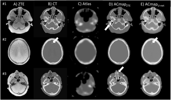
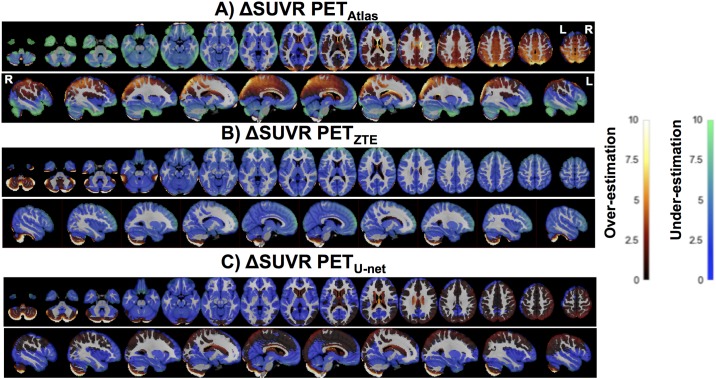
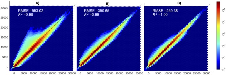
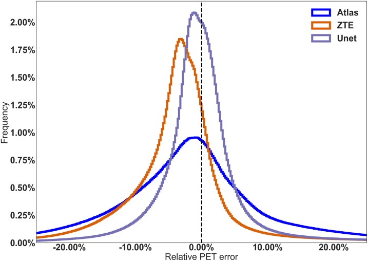
One of the main technical challenges of PET/MRI is to achieve an accurate PET attenuation correction (AC) estimation. In current systems, AC is accomplished by generating an MRI-based surrogate computed tomography (CT) from which AC-maps are derived. Nevertheless, all techniques currently implemented in clinical routine suffer from bias. We present here a convolutional neural network (CNN) that generated AC-maps from Zero Echo Time (ZTE) MR images. Seventy patients referred to our institution for 18FDG-PET/MR exam (SIGNA PET/MR, GE Healthcare) as part of the investigation of suspected dementia, were included. 23 patients were added to the training set of the manufacturer and 47 were used for validation. Brain computed tomography (CT) scan, two-point LAVA-flex MRI (for atlas-based AC) and ZTE-MRI were available in all patients. Three AC methods were evaluated and compared to CT-based AC (CTAC): one based on a single head-atlas, one based on ZTE-segmentation and one CNN with a 3D U-net architecture to generate AC maps from ZTE MR images. Impact on brain metabolism was evaluated combining voxel and regions-of-interest based analyses with CTAC set as reference. The U-net AC method yielded the lowest bias, the lowest inter-individual and inter-regional variability compared to PET images reconstructed with ZTE and Atlas methods. The impact on brain metabolism was negligible with average errors of -0.2% in most cortical regions. These results suggest that the U-net AC is more reliable for correcting photon attenuation in brain FDG-PET/MR than atlas-AC and ZTE-AC methods.
正电子发射断层磁共振成像(PET/MRI)的主要技术挑战之一是实现准确的 PET 衰减校正(AC)估计。在当前的系统中,通过生成基于 MRI 的替代计算机断层扫描(CT)来完成 AC,从中得出 AC 图。然而,目前在临床常规中实施的所有技术都存在偏差。我们在这里提出了一种卷积神经网络(CNN),它可以从零回波时间(ZTE)MR 图像生成 AC 图。我们纳入了 70 名因疑似痴呆而在我院接受 18FDG-PET/MR 检查(SIGNA PET/MR,GE Healthcare)的患者。23 名患者被添加到制造商的训练集中,47 名患者用于验证。所有患者均提供脑部 CT 扫描、两点 LAVA-flex MRI(用于基于图谱的 AC)和 ZTE-MRI。我们评估并比较了三种 AC 方法与基于 CT 的 AC(CTAC):一种基于单个头部图谱,一种基于 ZTE 分割,一种基于 3D U-net 架构的 CNN,用于从 ZTE MR 图像生成 AC 图。结合基于体素和感兴趣区域的分析,将 CTAC 作为参考,评估对脑代谢的影响。与使用 ZTE 和图谱方法重建的 PET 图像相比,U-net AC 方法的偏差最小,个体间和区域间的变异性最小。对大脑代谢的影响可以忽略不计,大多数皮质区域的平均误差为-0.2%。这些结果表明,与图谱-AC 和 ZTE-AC 方法相比,U-net AC 更可靠地校正脑 FDG-PET/MR 中的光子衰减。