• 文献检索
  • 文档翻译
  • 深度研究
  • 学术资讯
  • Suppr Zotero 插件Zotero 插件
  • 邀请有礼
  • 套餐&价格
  • 历史记录
应用&插件
Suppr Zotero 插件Zotero 插件浏览器插件Mac 客户端Windows 客户端微信小程序
定价
高级版会员购买积分包购买API积分包
服务
文献检索文档翻译深度研究API 文档MCP 服务
关于我们
关于 Suppr公司介绍联系我们用户协议隐私条款
关注我们

Suppr 超能文献

核心技术专利:CN118964589B侵权必究
粤ICP备2023148730 号-1Suppr @ 2026

文献检索

告别复杂PubMed语法,用中文像聊天一样搜索,搜遍4000万医学文献。AI智能推荐,让科研检索更轻松。

立即免费搜索

文件翻译

保留排版,准确专业,支持PDF/Word/PPT等文件格式,支持 12+语言互译。

免费翻译文档

深度研究

AI帮你快速写综述,25分钟生成高质量综述,智能提取关键信息,辅助科研写作。

立即免费体验

炎症细胞因子 IL-3 可能通过其蛋白 cargo 阻碍内皮细胞衍生的细胞外囊泡介导的心脏保护作用。

The Inflammatory Cytokine IL-3 Hampers Cardioprotection Mediated by Endothelial Cell-Derived Extracellular Vesicles Possibly via Their Protein Cargo.

机构信息

Department of Clinical and Biological Sciences, University of Turin, Regione Gonzole 10, 10043 Orbassano, Italy.

Department of Medical Sciences, University of Turin, Corso Dogliotti 14, 10126 Turin, Italy.

出版信息

Cells. 2020 Dec 23;10(1):13. doi: 10.3390/cells10010013.

DOI:10.3390/cells10010013
PMID:33374685
原文链接:https://pmc.ncbi.nlm.nih.gov/articles/PMC7822476/
Abstract

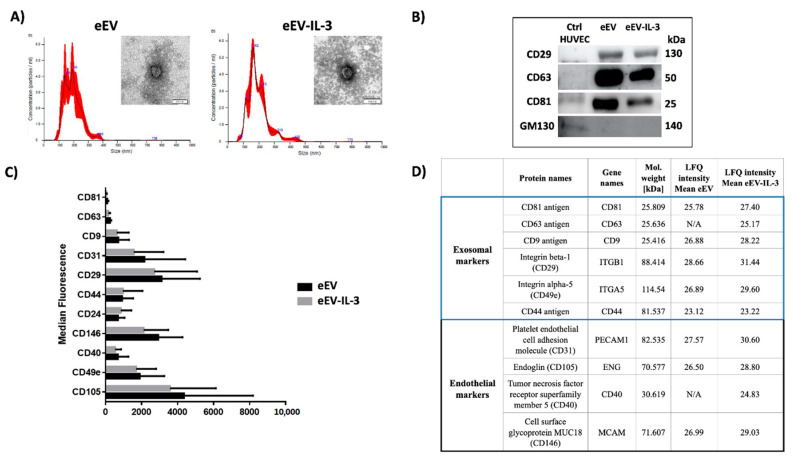
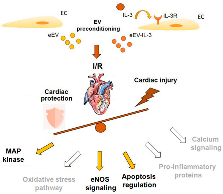
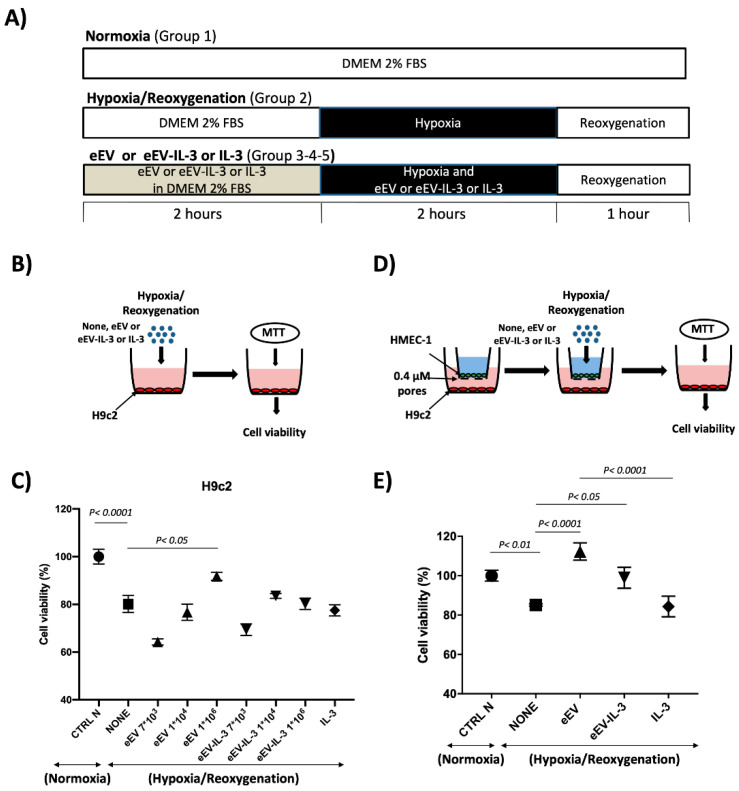
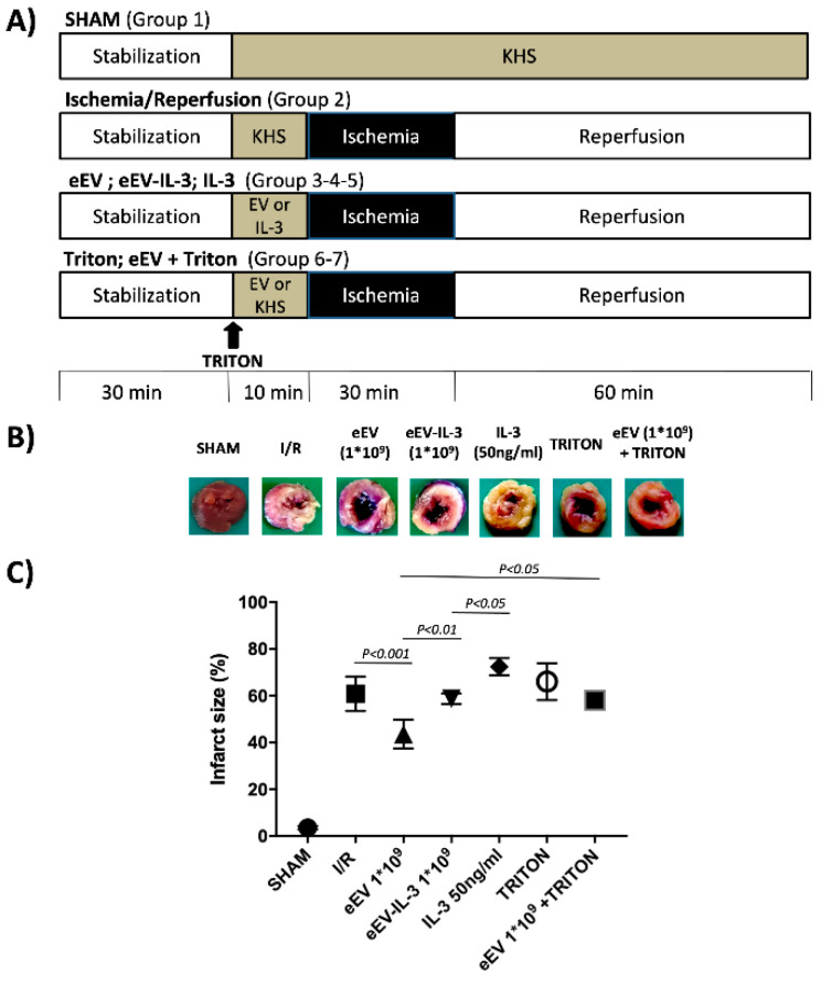
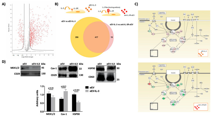
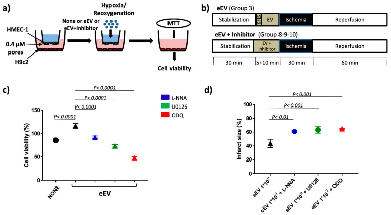
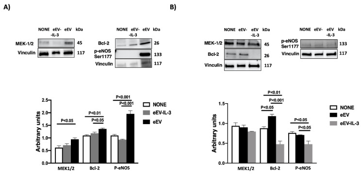
The biological relevance of extracellular vesicles (EV) released in an ischemia/reperfusion setting is still unclear. We hypothesized that the inflammatory microenvironment prevents cardioprotection mediated by endothelial cell (EC)-derived extracellular vesicles. The effects of naïve EC-derived EV (eEV) or eEV released in response to interleukin-3 (IL-3) (eEV-IL-3) were evaluated in cardiomyoblasts (H9c2) and rat hearts. In transwell assay, eEV protected the H9c2 exposed to hypoxia/reoxygenation (H/R) more efficiently than eEV-IL-3. Conversely, only eEV directly protected H9c2 cells to H/R-induced damage. Consistent with this latter observation, eEV, but not eEV-IL-3, exerted beneficial effects in the whole heart. Protein profiles of eEV and eEV-IL-3, established using label-free mass spectrometry, demonstrated that IL-3 drives changes in eEV-IL-3 protein cargo. Gene ontology analysis revealed that both eEV and eEV-IL-3 were equipped with full cardioprotective machinery, including the . eEV-IL-3 were also enriched in the endothelial-nitric oxide-synthase (eNOS)-antagonist caveolin-1 and proteins related to the inflammatory response. In vitro and ex vivo experiments demonstrated that a functional Mitogen-Activated Protein Kinase Kinase (MEK1/2)/eNOS/guanylyl-cyclase (GC) pathway is required for eEV-mediated cardioprotection. Consistently, eEV were found enriched in MEK1/2 and able to induce the expression of B-cell-lymphoma-2 (Bcl-2) and the phosphorylation of eNOS in vitro. We conclude that an inflammatory microenvironment containing IL-3 changes the eEV cargo and impairs eEV cardioprotective action.

摘要

细胞外囊泡(EV)在缺血/再灌注环境中释放的生物学相关性尚不清楚。我们假设炎症微环境会阻止内皮细胞(EC)衍生的细胞外囊泡介导的心脏保护作用。在心肌细胞(H9c2)和大鼠心脏中评估了幼稚 EC 衍生的 EV(eEV)或对白细胞介素 3(IL-3)反应释放的 eEV(eEV-IL-3)的作用。在 Transwell 测定中,eEV 比 eEV-IL-3更有效地保护暴露于缺氧/复氧(H/R)的 H9c2。相反,只有 eEV 直接保护 H9c2 细胞免受 H/R 诱导的损伤。与后一种观察结果一致,eEV 而不是 eEV-IL-3 在整个心脏中发挥有益作用。使用无标记质谱法建立的 eEV 和 eEV-IL-3 的蛋白质谱表明,IL-3 驱动 eEV-IL-3 蛋白货物的变化。GO 分析表明,eEV 和 eEV-IL-3 都配备了完整的心脏保护机制,包括. eEV-IL-3 还富含内皮一氧化氮合酶(eNOS)拮抗剂 caveolin-1 和与炎症反应相关的蛋白质。体外和离体实验表明,功能性丝裂原激活蛋白激酶激酶(MEK1/2)/eNOS/鸟苷酸环化酶(GC)途径对于 eEV 介导的心脏保护作用是必需的。一致地,发现 eEV 富含 MEK1/2,并能够在体外诱导 B 细胞淋巴瘤-2(Bcl-2)的表达和 eNOS 的磷酸化。我们得出结论,含有 IL-3 的炎症微环境改变了 eEV 的货物,并损害了 eEV 的心脏保护作用。

https://cdn.ncbi.nlm.nih.gov/pmc/blobs/1242/7822476/6bb47feedc57/cells-10-00013-g007.jpg
https://cdn.ncbi.nlm.nih.gov/pmc/blobs/1242/7822476/eb91da69f7f2/cells-10-00013-g001.jpg
https://cdn.ncbi.nlm.nih.gov/pmc/blobs/1242/7822476/4b478156bee0/cells-10-00013-g002.jpg
https://cdn.ncbi.nlm.nih.gov/pmc/blobs/1242/7822476/9f8735121c8b/cells-10-00013-g003.jpg
https://cdn.ncbi.nlm.nih.gov/pmc/blobs/1242/7822476/aadd15ac2bc4/cells-10-00013-g004.jpg
https://cdn.ncbi.nlm.nih.gov/pmc/blobs/1242/7822476/8d516e39ff65/cells-10-00013-g005.jpg
https://cdn.ncbi.nlm.nih.gov/pmc/blobs/1242/7822476/8520bea3d7a8/cells-10-00013-g006.jpg
https://cdn.ncbi.nlm.nih.gov/pmc/blobs/1242/7822476/6bb47feedc57/cells-10-00013-g007.jpg

相似文献

1
The Inflammatory Cytokine IL-3 Hampers Cardioprotection Mediated by Endothelial Cell-Derived Extracellular Vesicles Possibly via Their Protein Cargo.炎症细胞因子 IL-3 可能通过其蛋白 cargo 阻碍内皮细胞衍生的细胞外囊泡介导的心脏保护作用。
Cells. 2020 Dec 23;10(1):13. doi: 10.3390/cells10010013.
2
Cardiomyocyte-derived small extracellular vesicles can signal eNOS activation in cardiac microvascular endothelial cells to protect against Ischemia/Reperfusion injury.心肌细胞衍生的小细胞外囊泡可在心脏微血管内皮细胞中发出信号,激活 eNOS,从而防止缺血/再灌注损伤。
Theranostics. 2020 Sep 23;10(25):11754-11774. doi: 10.7150/thno.43163. eCollection 2020.
3
Emodin, A Chinese Herbal Medicine, Inhibits Reoxygenation-Induced Injury in Cultured Human Aortic Endothelial Cells by Regulating the Peroxisome Proliferator-Activated Receptor-γ (PPAR-γ) and Endothelial Nitric Oxide Synthase (eNOS) Signaling Pathway.大黄素,一种中药,通过调节过氧化物酶体增殖物激活受体-γ(PPAR-γ)和内皮型一氧化氮合酶(eNOS)信号通路抑制培养的人主动脉内皮细胞再氧合诱导的损伤。
Med Sci Monit. 2018 Feb 1;24:643-651. doi: 10.12659/msm.908237.
4
Glucose and angiotensin II-derived endothelial extracellular vesicles regulate endothelial dysfunction via ERK1/2 activation.葡萄糖和血管紧张素II衍生的内皮细胞外囊泡通过激活ERK1/2调节内皮功能障碍。
Pflugers Arch. 2017 Feb;469(2):293-302. doi: 10.1007/s00424-016-1926-2. Epub 2016 Dec 14.
5
Interleukin-6 inhibits endothelial nitric oxide synthase activation and increases endothelial nitric oxide synthase binding to stabilized caveolin-1 in human vascular endothelial cells.白细胞介素-6 抑制内皮型一氧化氮合酶的激活,并增加人血管内皮细胞中内皮型一氧化氮合酶与稳定化 caveolin-1 的结合。
J Hypertens. 2010 May;28(5):940-51. doi: 10.1097/HJH.0b013e32833992ef.
6
Extracellular Vesicles Work as a Functional Inflammatory Mediator Between Vascular Endothelial Cells and Immune Cells.细胞外囊泡在血管内皮细胞和免疫细胞之间充当功能性炎症介质。
Front Immunol. 2018 Aug 6;9:1789. doi: 10.3389/fimmu.2018.01789. eCollection 2018.
7
Endothelial extracellular vesicles contain protective proteins and rescue ischemia-reperfusion injury in a human heart-on-chip.内皮细胞外囊泡含有保护性蛋白,并能在人心脏芯片中挽救缺血再灌注损伤。
Sci Transl Med. 2020 Oct 14;12(565). doi: 10.1126/scitranslmed.aax8005.
8
Activated Stat5 trafficking Via Endothelial Cell-derived Extracellular Vesicles Controls IL-3 Pro-angiogenic Paracrine Action.通过内皮细胞衍生的细胞外囊泡进行的活化Stat5转运控制IL-3促血管生成旁分泌作用。
Sci Rep. 2016 May 9;6:25689. doi: 10.1038/srep25689.
9
Cardioprotection of CAPE-oNO against myocardial ischemia/reperfusion induced ROS generation via regulating the SIRT1/eNOS/NF-κB pathway in vivo and in vitro.CAPE-oNO 通过体内和体外调节 SIRT1/eNOS/NF-κB 通路对心肌缺血/再灌注诱导的 ROS 生成的心脏保护作用。
Redox Biol. 2018 May;15:62-73. doi: 10.1016/j.redox.2017.11.023. Epub 2017 Nov 29.
10
Key role of endothelium in the eNOS-dependent cardioprotection with exercise training.内皮在运动训练介导的eNOS依赖性心脏保护中的关键作用。
J Mol Cell Cardiol. 2017 Jan;102:26-30. doi: 10.1016/j.yjmcc.2016.11.008. Epub 2016 Nov 17.

引用本文的文献

1
Cardioprotection Reloaded: Reflections on 40 Years of Research.心脏保护重装上阵:40年研究回顾
Antioxidants (Basel). 2025 Jul 18;14(7):889. doi: 10.3390/antiox14070889.
2
Research progress of exosomes from different sources in myocardial ischemia.不同来源外泌体在心肌缺血中的研究进展
Front Cardiovasc Med. 2024 Sep 12;11:1436764. doi: 10.3389/fcvm.2024.1436764. eCollection 2024.
3
Identifying plasma proteomic signatures from health to heart failure, across the ejection fraction spectrum.从健康到心力衰竭,跨越射血分数谱识别血浆蛋白质组学特征。

本文引用的文献

1
The Contrasting Role of Extracellular Vesicles in Vascular Inflammation and Tissue Repair.细胞外囊泡在血管炎症和组织修复中的对比作用
Front Pharmacol. 2019 Dec 17;10:1479. doi: 10.3389/fphar.2019.01479. eCollection 2019.
2
Ischemia Reperfusion Injury: Mechanisms of Damage/Protection and Novel Strategies for Cardiac Recovery/Regeneration.缺血再灌注损伤:损伤/保护机制和心脏恢复/再生的新策略。
Int J Mol Sci. 2019 Oct 11;20(20):5024. doi: 10.3390/ijms20205024.
3
α-Cyclodextrin and α-Cyclodextrin Polymers as Oxygen Nanocarriers to Limit Hypoxia/Reoxygenation Injury: Implications from an In Vitro Model.
Sci Rep. 2024 Jun 27;14(1):14871. doi: 10.1038/s41598-024-65667-0.
4
High placental expression of FLT1, LEP, PHYHIP and IL3RA - In persons of African ancestry with severe preeclampsia.在非洲裔严重先兆子痫患者中,胎盘高表达 FLT1、LEP、PHYHIP 和 IL3RA。
Placenta. 2023 Dec;144:13-22. doi: 10.1016/j.placenta.2023.10.008. Epub 2023 Oct 20.
5
Atorvastatin-pretreated mesenchymal stem cell-derived extracellular vesicles promote cardiac repair after myocardial infarction via shifting macrophage polarization by targeting microRNA-139-3p/Stat1 pathway.阿托伐他汀预处理的间充质干细胞衍生的细胞外囊泡通过靶向 microRNA-139-3p/Stat1 通路转移巨噬细胞极化促进心肌梗死后的心脏修复。
BMC Med. 2023 Mar 16;21(1):96. doi: 10.1186/s12916-023-02778-x.
6
Pathways for Cardioprotection in Perspective: Focus on Remote Conditioning and Extracellular Vesicles.心脏保护途径展望:聚焦远程预处理与细胞外囊泡
Biology (Basel). 2023 Feb 14;12(2):308. doi: 10.3390/biology12020308.
7
The Influence of Comorbidities on Chemokine and Cytokine Profile in Obstructive Sleep Apnea Patients: Preliminary Results.合并症对阻塞性睡眠呼吸暂停患者趋化因子和细胞因子谱的影响:初步结果
J Clin Med. 2023 Jan 19;12(3):801. doi: 10.3390/jcm12030801.
8
Extracellular vesicles in cardiac repair and regeneration: Beyond stem-cell-based approaches.心脏修复与再生中的细胞外囊泡:超越基于干细胞的方法
Front Cell Dev Biol. 2022 Sep 2;10:996887. doi: 10.3389/fcell.2022.996887. eCollection 2022.
9
Lymphatic and Blood Endothelial Extracellular Vesicles: A Story Yet to Be Written.淋巴和血液内皮细胞外囊泡:一个有待书写的故事。
Life (Basel). 2022 Apr 28;12(5):654. doi: 10.3390/life12050654.
10
Plasma-derived extracellular vesicles transfer microRNA-130a-3p to alleviate myocardial ischemia/reperfusion injury by targeting ATG16L1.血浆衍生的细胞外囊泡通过靶向 ATG16L1 转移 microRNA-130a-3p 来减轻心肌缺血/再灌注损伤。
Cell Tissue Res. 2022 Jul;389(1):99-114. doi: 10.1007/s00441-022-03605-0. Epub 2022 May 3.
α-环糊精及其聚合物作为氧纳米载体限制缺氧/复氧损伤:来自体外模型的启示
Polymers (Basel). 2018 Feb 22;10(2):211. doi: 10.3390/polym10020211.
4
Coronary Microvascular Injury in Reperfused Acute Myocardial Infarction: A View From an Integrative Perspective.再灌注急性心肌梗死中的冠状动脉微血管损伤:综合视角下的观点
J Am Heart Assoc. 2018 Nov 6;7(21):e009949. doi: 10.1161/JAHA.118.009949.
5
Specificities of secretion and uptake of exosomes and other extracellular vesicles for cell-to-cell communication.细胞间通讯中外泌体和其他细胞外囊泡的分泌和摄取的特异性。
Nat Cell Biol. 2019 Jan;21(1):9-17. doi: 10.1038/s41556-018-0250-9. Epub 2019 Jan 2.
6
Effects of tumor necrosis factor-α-induced exosomes on the endothelial cellular behavior, metabolism and bioenergetics.肿瘤坏死因子-α诱导的外泌体对内皮细胞行为、代谢和生物能量学的影响。
Microcirculation. 2019 Jan;26(1):e12515. doi: 10.1111/micc.12515. Epub 2018 Nov 29.
7
Endothelial cells release cardioprotective exosomes that may contribute to ischaemic preconditioning.内皮细胞释放具有心脏保护作用的外泌体,可能有助于缺血预处理。
Sci Rep. 2018 Oct 26;8(1):15885. doi: 10.1038/s41598-018-34357-z.
8
Practical guidelines for rigor and reproducibility in preclinical and clinical studies on cardioprotection.心脏保护临床前和临床研究中严谨性与可重复性的实用指南。
Basic Res Cardiol. 2018 Aug 17;113(5):39. doi: 10.1007/s00395-018-0696-8.
9
Cardioprotective Properties of Human Platelets Are Lost in Uncontrolled Diabetes Mellitus: A Study in Isolated Rat Hearts.人血小板的心脏保护特性在未控制的糖尿病中丧失:对离体大鼠心脏的一项研究。
Front Physiol. 2018 Jul 10;9:875. doi: 10.3389/fphys.2018.00875. eCollection 2018.
10
Protective effects of circulating microvesicles derived from ischemic preconditioning on myocardial ischemia/reperfusion injury in rats by inhibiting endoplasmic reticulum stress.循环微小泡源自缺血预处理对大鼠心肌缺血/再灌注损伤的保护作用,通过抑制内质网应激。
Apoptosis. 2018 Aug;23(7-8):436-448. doi: 10.1007/s10495-018-1469-4.