Radiation Oncology Branch, Center for Cancer Research, National Cancer Institute, National Institutes of Health, Bethesda, Maryland.
Experimental Pathology Laboratory, Laboratory of Pathology, Center for Cancer Research, National Cancer Institute, National Institutes of Health, Bethesda, Maryland; Department of Pathology, Asan Medical Center, University of Ulsan College of Medicine, Seoul, Republic of Korea.
Int J Radiat Oncol Biol Phys. 2021 Jun 1;110(2):526-538. doi: 10.1016/j.ijrobp.2020.12.035. Epub 2020 Dec 30.
Type II pneumocyte (alveolar epithelial cells type II [AECII]) senescence has been implicated in the progression of lung fibrosis. The capacity of senescent cells to modulate pulmonary macrophages to drive fibrosis is unexplored. Insulin-like growth factor-1 receptor (IGF-1R) signaling has been implicated as a regulator of senescence and aging.
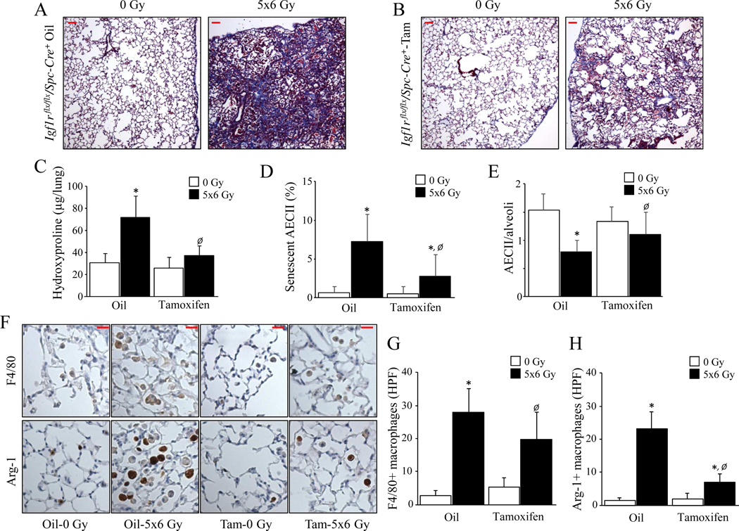
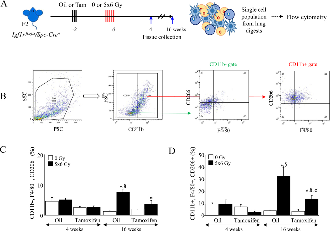
Mice with an AECII-specific deletion of IGF-1R received thoracic irradiation (n ≥ 5 per condition), and the effect of IGF-1R deficiency on radiation-induced AECII senescence and macrophage polarization to an alternatively activated phenotype (M2) was investigated. IGF-1R signaling, macrophage polarization, and senescence were evaluated in surgically resected human lung (n = 63).
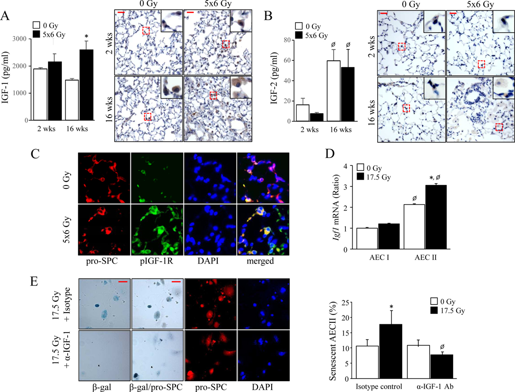
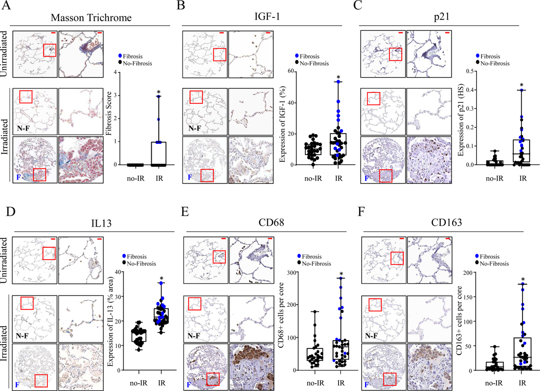
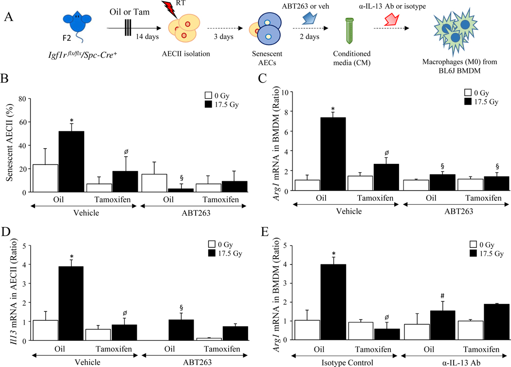
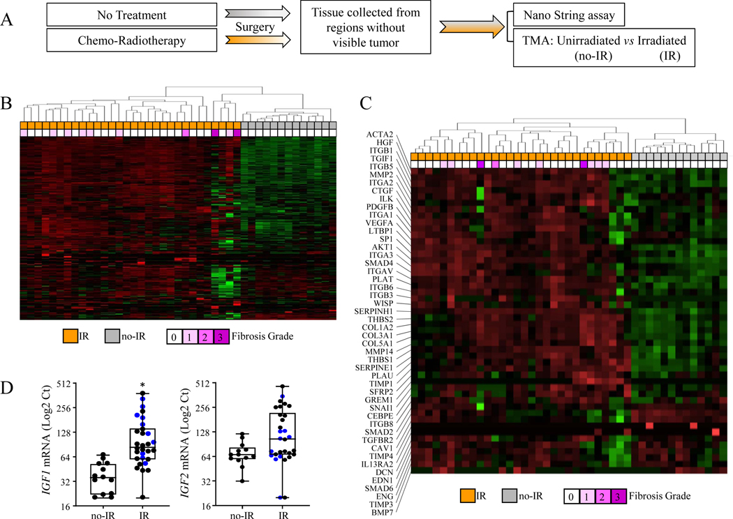
IGF-1R deficient mice demonstrated reduced AECII senescence (senescent AECII/field; intact: 7.25% ± 3.5% [mean ± SD], deficient: 2.75% ± 2.8%, P = .0001), reduced accumulation of M2 macrophages (intact: 24.7 ± 2.2 cells/field, deficient: 15.5 ± 1.2 cells/field, P = .0086), and fibrosis (hydroxyproline content; intact: 71.9 ± 21.7 μg/lung, deficient: 31.7 ± 7.9, P = .0485) after irradiation. Senescent AECII enhanced M2 polarization in a paracrine fashion (relative Arg1 mRNA, 0 Gy: 1.0 ± 0.4, 17.5 Gy: 7.34 ± 0.5, P < .0001). Evaluation of surgical samples from patients treated with chemoradiation demonstrated increased expression of IGF-1 (unirradiated: 10.2% ± 4.9% area, irradiated: 15.1% ± 11.5%, P = .0377), p21 (unirradiated: 0.013 ± 0.02 histoscore, irradiated: 0.084 ± 0.09 histoscore, P = .0002), IL-13 (unirradiated: 13.7% ± 2.8% area, irradiated: 21.7% ± 3.8%, P < .0001), and M2 macrophages in fibrotic regions relative to nonfibrotic regions (unirradiated: 11.4 ± 12.2 CD163 + cells/core, irradiated: 43.1 ± 40.9 cells/core, P = .0011), consistent with findings from animal models of lung fibrosis.
This study demonstrates that senescent AECII are necessary for the progression of pulmonary fibrosis and serve as a targetable, chronic stimuli for macrophage activation in fibrotic lung.
Ⅱ型肺泡上皮细胞(肺泡上皮细胞Ⅱ型 [AECII])衰老与肺纤维化的进展有关。衰老细胞调节肺巨噬细胞向纤维化表型(M2)极化的能力尚未被探索。胰岛素样生长因子-1 受体(IGF-1R)信号已被认为是衰老和老化的调节剂。
接受 AECII 特异性 IGF-1R 缺失的小鼠接受了胸部照射(每组≥5 只),并研究了 IGF-1R 缺失对辐射诱导的 AECII 衰老和巨噬细胞向替代激活表型(M2)极化的影响。在手术切除的人类肺中评估 IGF-1R 信号、巨噬细胞极化和衰老(n = 63)。
IGF-1R 缺失的小鼠表现出 AECII 衰老减少(完整:7.25%±3.5%,缺陷:2.75%±2.8%,P =.0001),M2 巨噬细胞积聚减少(完整:24.7±2.2 个/场,缺陷:15.5±1.2 个/场,P =.0086),照射后纤维化(羟脯氨酸含量;完整:71.9±21.7μg/肺,缺陷:31.7±7.9μg/肺,P =.0485)。衰老的 AECII 以旁分泌方式增强 M2 极化(相对 Arg1 mRNA,0 Gy:1.0±0.4,17.5 Gy:7.34±0.5,P <.0001)。对接受放化疗的患者手术样本的评估显示 IGF-1 表达增加(未照射:10.2%±4.9%面积,照射:15.1%±11.5%,P =.0377),p21(未照射:0.013±0.02 组织评分,照射:0.084±0.09 组织评分,P =.0002),IL-13(未照射:13.7%±2.8%面积,照射:21.7%±3.8%,P <.0001)和 M2 巨噬细胞在纤维化区域相对于非纤维化区域(未照射:11.4±12.2 CD163 +细胞/核,照射:43.1±40.9 细胞/核,P =.0011),与肺纤维化动物模型的发现一致。
本研究表明,衰老的 AECII 是肺纤维化进展所必需的,并且是纤维化肺中巨噬细胞激活的可靶向慢性刺激物。