Achucarro Basque Center for Neuroscience, 48940 Leioa, Spain.
CIC biomaGUNE, Basque Research and Technology Alliance (BRTA), Paseo Miramon 182, 20014, San Sebastian, Spain.
Theranostics. 2021 Jan 1;11(1):410-425. doi: 10.7150/thno.51046. eCollection 2021.
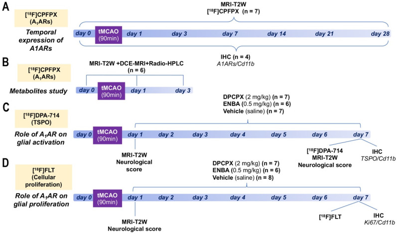
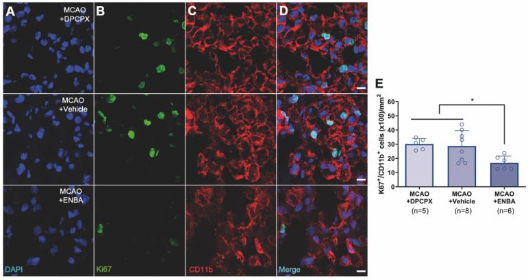
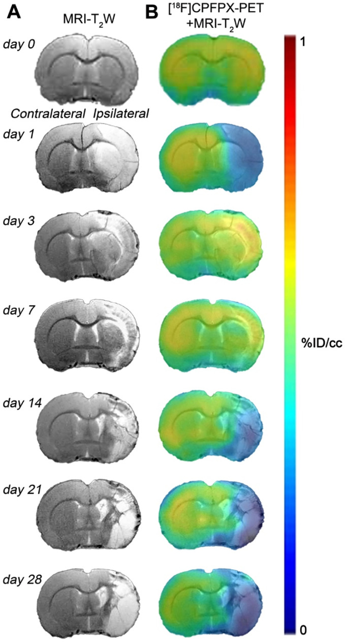
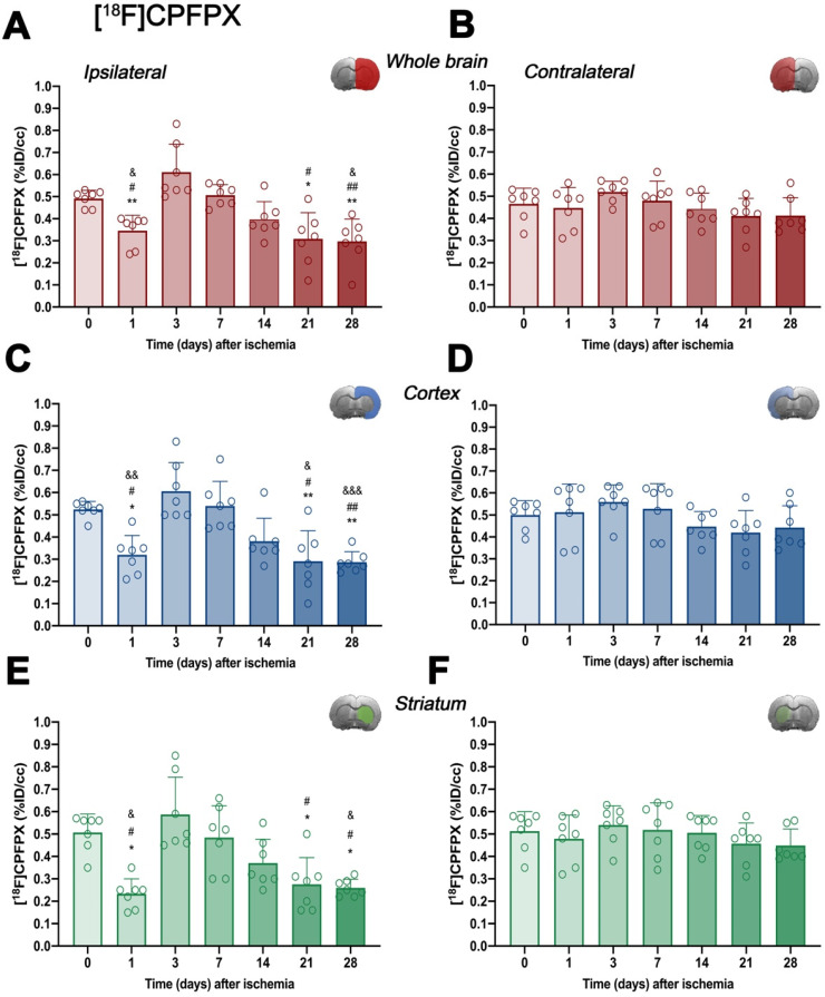
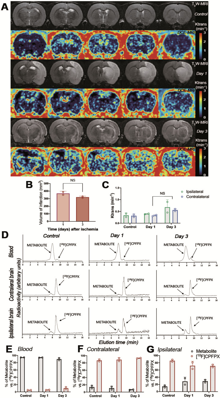
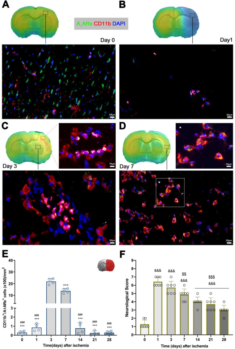
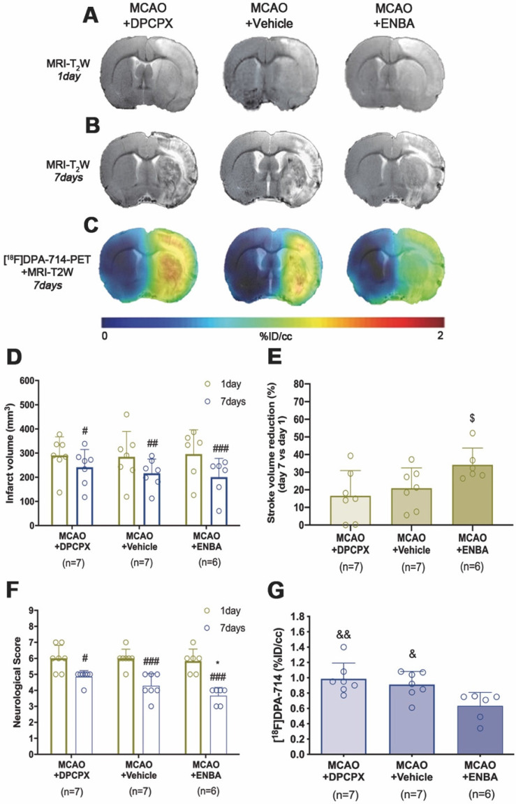
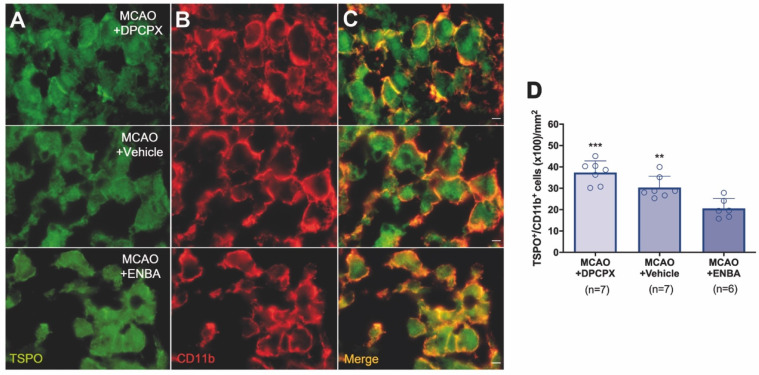
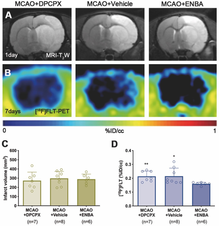
Adenosine A receptors (AARs) are promising imaging biomarkers and targets for the treatment of stroke. Nevertheless, the role of AARs on ischemic damage and its subsequent neuroinflammatory response has been scarcely explored so far. In this study, the expression of AARs after transient middle cerebral artery occlusion (MCAO) was evaluated by positron emission tomography (PET) with [F]CPFPX and immunohistochemistry (IHC). In addition, the role of AARs on stroke inflammation using pharmacological modulation was assessed with magnetic resonance imaging (MRI), PET imaging with [F]DPA-714 (TSPO) and [F]FLT (cellular proliferation), as well as IHC and neurofunctional studies. In the ischemic territory, [F]CPFPX signal and IHC showed the overexpression of AARs in microglia and infiltrated leukocytes after cerebral ischemia. Ischemic rats treated with the AAR agonist ENBA showed a significant decrease in both [F]DPA-714 and [F]FLT signal intensities at day 7 after cerebral ischemia, a feature that was confirmed by IHC results. Besides, the activation of AARs promoted the reduction of the brain lesion, as measured with TW-MRI, and the improvement of neurological outcome including motor, sensory and reflex responses. These results show for the first time the PET imaging of AARs expression after cerebral ischemia in rats and the application of [F]FLT to evaluate glial proliferation in response to treatment. Notably, these data provide evidence for AARs playing a key role in the control of both the activation of resident glia and the proliferation of microglia and macrophages after experimental stroke in rats.
腺苷 A 受体 (AARs) 是有前途的成像生物标志物和治疗中风的靶点。然而,到目前为止,AARs 在缺血性损伤及其随后的神经炎症反应中的作用还鲜有研究。在这项研究中,通过正电子发射断层扫描 (PET) 用 [F]CPFPX 和免疫组织化学 (IHC) 评估短暂性大脑中动脉闭塞 (MCAO) 后 AARs 的表达。此外,还通过磁共振成像 (MRI)、用 [F]DPA-714(TSPO)和 [F]FLT(细胞增殖)进行 PET 成像以及 IHC 和神经功能研究,评估了 AARs 对中风炎症的作用。在缺血区域,[F]CPFPX 信号和 IHC 显示在脑缺血后小胶质细胞和浸润的白细胞中 AARs 过度表达。用 AAR 激动剂 ENBA 治疗的缺血大鼠在脑缺血后 7 天,[F]DPA-714 和 [F]FLT 信号强度均显著降低,这一特征通过 IHC 结果得到证实。此外,AARs 的激活促进了 TW-MRI 测量的脑损伤减少,以及运动、感觉和反射反应等神经功能的改善。这些结果首次显示了大鼠脑缺血后 AARs 表达的 PET 成像,以及用 [F]FLT 评估对治疗的反应性胶质细胞增殖。值得注意的是,这些数据为 AARs 在控制实验性中风后驻留胶质细胞的激活和小胶质细胞和巨噬细胞的增殖方面发挥关键作用提供了证据。