Tianjin Key Laboratory of Neurotrauma Repair, Institute of Traumatic Brain Injury and Neuroscience, Center for Neurology and Neurosurgery of Characteristic Medical Center of Chinese People's Armed Police Force (PAP), Chenglin Road No.220, Tianjin 300162, China.
Postgraduate school, Medical school of Chinese People's Liberation Army (PLA), General Hospital of PLA, Fuxing Road No. 28, Beijing 100853, China.
Theranostics. 2021 Jan 1;11(2):768-788. doi: 10.7150/thno.50540. eCollection 2021.
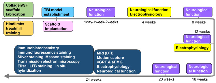
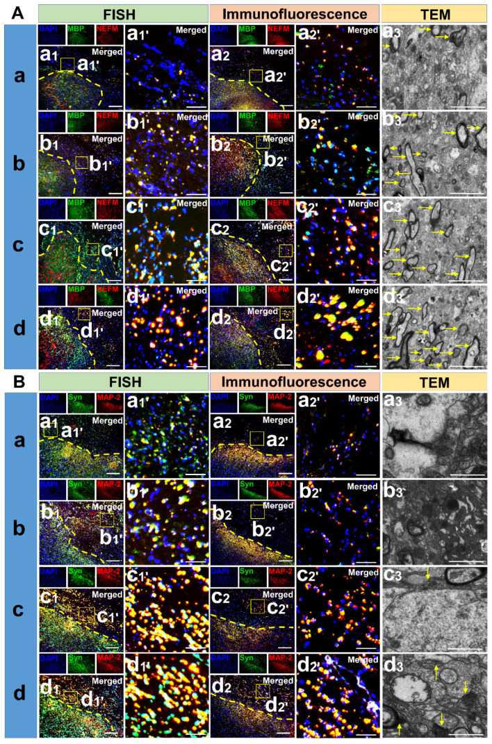
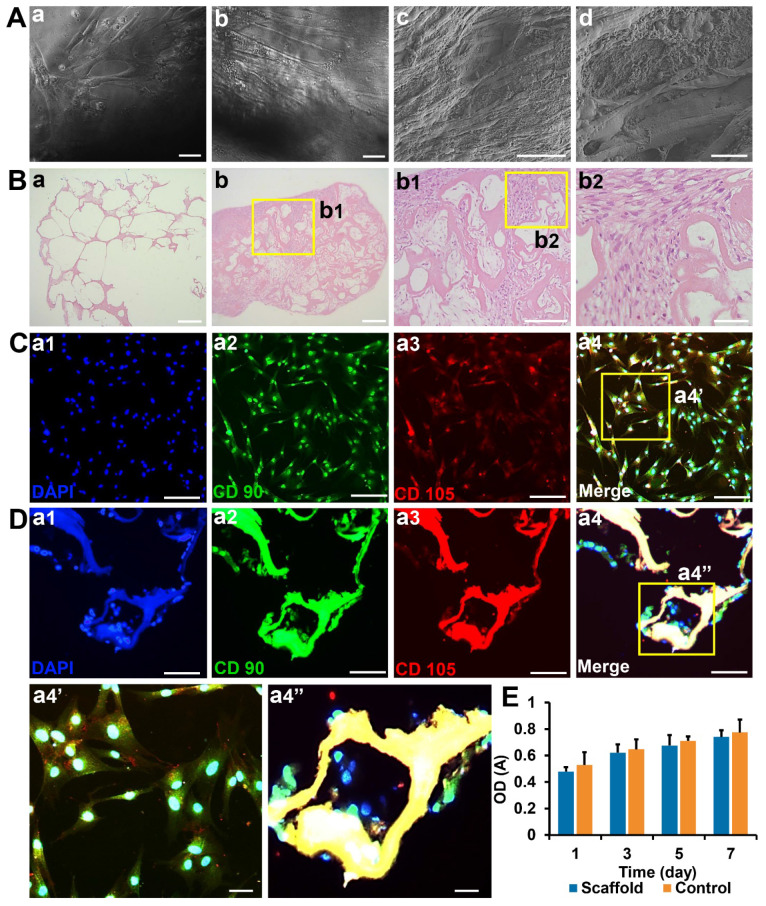
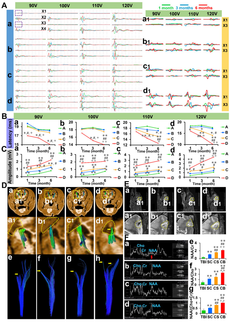
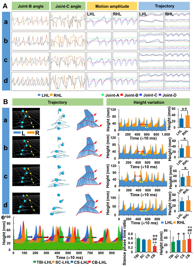
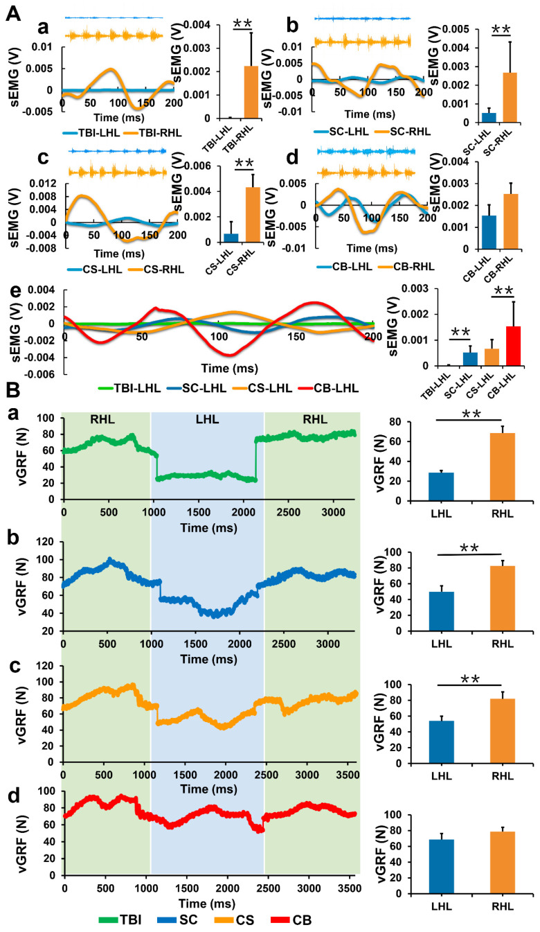
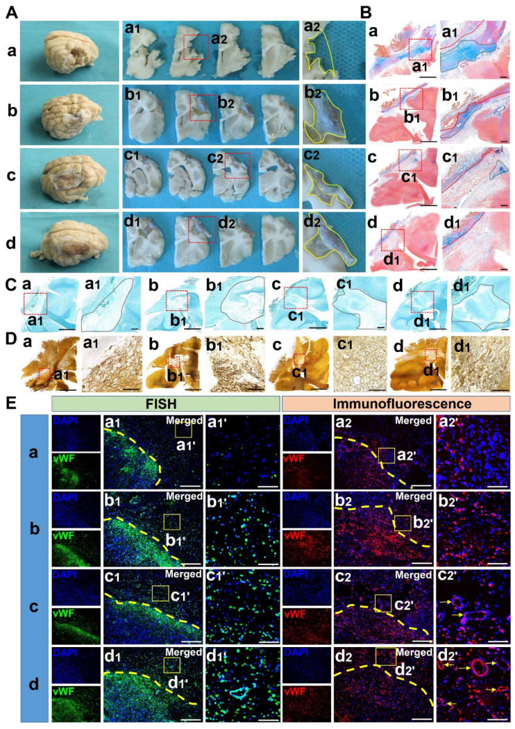
The combination of medical and tissue engineering in neural regeneration studies is a promising field. Collagen, silk fibroin and seed cells are suitable options and have been widely used in the repair of spinal cord injury. In this study, we aimed to determine whether the implantation of a complex fabricated with collagen/silk fibroin (SF) and the human umbilical cord mesenchymal stem cells (hUCMSCs) can promote cerebral cortex repair and motor functional recovery in a canine model of traumatic brain injury (TBI). A porous scaffold was fabricated with cross-linked collagen and SF. Its physical properties and degeneration rate were measured. The scaffolds were co-cultured with hUCMSCs after which an implantable complex was formed. After complex implantation to a canine model of TBI, the motor evoked potential (MEP) and magnetic resonance imaging (MRI) were used to evaluate the integrity of the cerebral cortex. The neurologic score, motion capture, surface electromyography (sEMG), and vertical ground reaction force (vGRF) were measured in the analysis of motor functions. In vitro analysis of inflammation levels was performed by Elisa while immunohistochemistry was used in track the fate of hUCMSCs. In situ hybridization, transmission electron microscope, and immunofluorescence were used to assess neural and vascular regeneration. Favorable physical properties, suitable degradation rate, and biocompatibility were observed in the collagen/SF scaffolds. The group with complex implantation exhibited the best cerebral cortex integrity and motor functions. The implantation also led to the regeneration of more blood vessels and nerve fibers, less glial fibers, and inflammatory factors. Implantation of this complex enhanced therapy in traumatic brain injury (TBI) through structural repair and functional recovery. These effects exhibit the translational prospects for the clinical application of this complex.
在神经再生研究中,医学与组织工程学的结合是一个很有前景的领域。胶原蛋白、丝素纤维和种子细胞是合适的选择,已广泛应用于脊髓损伤的修复。在这项研究中,我们旨在确定胶原蛋白/丝素纤维(SF)和人脐带间充质干细胞(hUCMSCs)的复合物的植入是否可以促进创伤性脑损伤(TBI)犬模型的大脑皮质修复和运动功能恢复。用交联的胶原蛋白和 SF 制造了一种多孔支架。测量了其物理性能和降解率。支架与 hUCMSCs 共培养后,形成了可植入的复合物。在 TBI 犬模型中植入复合物后,使用运动诱发电位(MEP)和磁共振成像(MRI)评估大脑皮质的完整性。通过神经评分、运动捕捉、表面肌电图(sEMG)和垂直地面反力(vGRF)分析运动功能。通过 Elisa 进行体外炎症水平分析,并用免疫组织化学跟踪 hUCMSCs 的命运。原位杂交、透射电镜和免疫荧光用于评估神经和血管再生。胶原蛋白/SF 支架具有良好的物理性能、适宜的降解率和生物相容性。复合物植入组表现出最佳的大脑皮质完整性和运动功能。植入物还导致更多的血管和神经纤维再生,更少的神经胶质纤维和炎症因子。这种复合物的植入通过结构修复和功能恢复增强了创伤性脑损伤(TBI)的治疗效果。这些效果展示了该复合物临床应用的转化前景。