Suh Soh Youn, Le Alan, Shin Andrew, Park Joseph, Demer Joseph L
Department of Ophthalmology, University of California, Los Angeles, California, United States.
Department of Neuroengineering, University of California, Los Angeles, California, United States.
Invest Ophthalmol Vis Sci. 2017 Oct 1;58(12):5015-5021. doi: 10.1167/iovs.17-22596.
We investigated the effect of graded range of horizontal duction on the shape of the peripapillary Bruch's membrane (ppBM) and optic nerve head (ONH).
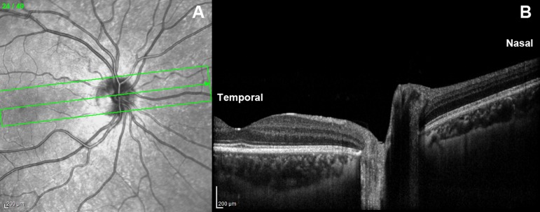
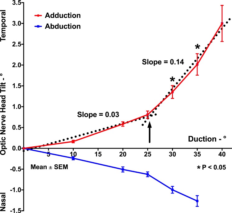
In 50 eyes of 25 normal subjects, the ONH and peripapillary retina were imaged by optical coherence tomography (OCT) in central gaze and incremental angles of add- and abduction. Displacements of the Bruch's membrane opening (BMO), optic cup (OC), and change in ONH angle in eccentric gazes were compared to those of central gaze, in add- and abduction.
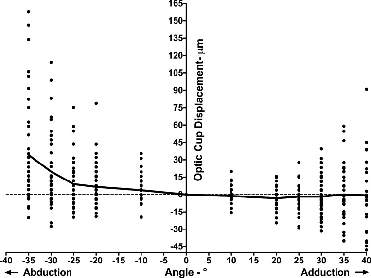
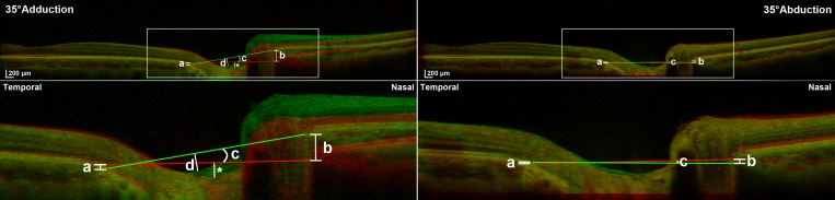
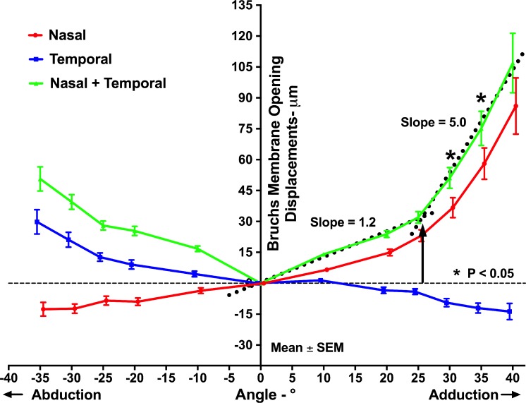
With increasing duction, the nasal edge of the BMO (nBMO) shifted progressively anteriorly in adduction and posteriorly in abduction, while the temporal edge of the BMO (tBMO) shifted posteriorly in adduction and anteriorly in abduction. The summed absolute nBMO and tBMO displacements in 30° and 35° adduction significantly exceeded those in comparable abduction angles (P < 0.005 for both). The ONH progressively tilted temporally in adduction and nasally in abduction; absolute ONH tilt in adduction was significantly greater than that in abduction for 30° and 35° ductions (P < 0.005 for both). BMO displacement and ONH tilt in adduction exhibited bilinear behavior, with greater effects for both at angles exceeding 26°. The OC shifted significantly farther anteriorly in abduction than adduction at every angle from 10° to 35°.
Horizontal duction deforms the ONH and ppBM, but more in adduction than in abduction, and increasingly so for angles greater than 26°. This behavior is consistent with optic nerve sheath tethering for adduction exceeding 26°.
我们研究了水平眼外肌运动的分级范围对视乳头周围 Bruch 膜(ppBM)和视神经乳头(ONH)形状的影响。
对 25 名正常受试者的 50 只眼睛,采用光学相干断层扫描(OCT)在中心注视以及内收和外展的递增角度下对视神经乳头和视乳头周围视网膜进行成像。比较内收和外展时偏心注视下 Bruch 膜开口(BMO)、视杯(OC)的位移以及视神经乳头角度的变化与中心注视时的情况。
随着眼外肌运动增加,BMO 的鼻侧边缘(nBMO)在内收时逐渐向前移位,在外展时向后移位,而 BMO 的颞侧边缘(tBMO)在内收时向后移位,在外展时向前移位。在 30°和 35°内收时,nBMO 和 tBMO 的绝对位移总和显著超过可比外展角度时的位移总和(两者 P < 0.005)。视神经乳头在内收时逐渐向颞侧倾斜,在外展时向鼻侧倾斜;在 30°和 35°眼外肌运动时,内收时视神经乳头的绝对倾斜度显著大于外展时(两者 P < 0.005)。内收时 BMO 位移和视神经乳头倾斜呈现双线性行为,在超过 26°的角度时两者影响更大。在从 10°到 35°的每个角度,视杯在外展时向前移位明显比内收时更远。
水平眼外肌运动使视神经乳头和 ppBM 变形,但内收时比外展时更明显,且在大于 26°的角度时愈发如此。这种行为与内收超过 26°时视神经鞘的束缚作用一致。