Biomedical Photonic Imaging (BMPI), Technical Medical Center, University of Twente, 7500 AE Enschede, The Netherlands.
Research & Business Development Division, CYBERDYNE Inc., Cambridge Innovation Center, 3013 AK Rotterdam, The Netherlands.
Sensors (Basel). 2021 Jan 4;21(1):283. doi: 10.3390/s21010283.
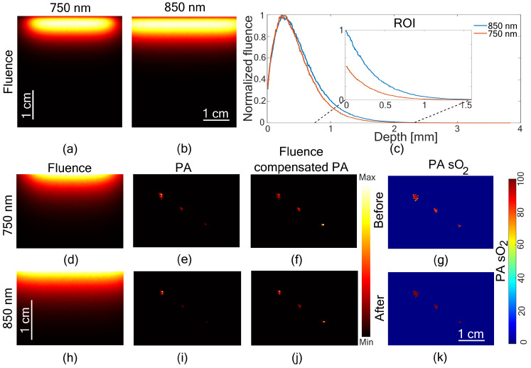
Oxygen saturation imaging has potential in several preclinical and clinical applications. Dual-wavelength LED array-based photoacoustic oxygen saturation imaging can be an affordable solution in this case. For the translation of this technology, there is a need to improve its accuracy and validate it against ground truth methods. We propose a fluence compensated oxygen saturation imaging method, utilizing structural information from the ultrasound image, and prior knowledge of the optical properties of the tissue with a Monte-Carlo based light propagation model for the dual-wavelength LED array configuration. We then validate the proposed method with oximeter measurements in tissue-mimicking phantoms. Further, we demonstrate in vivo imaging on small animal and a human subject. We conclude that the proposed oxygen saturation imaging can be used to image tissue at a depth of 6-8 mm in both preclinical and clinical applications.
氧饱和度成像是几种临床前和临床应用中的潜在手段。基于双波长 LED 阵列的光声氧饱和度成像在这种情况下可能是一种经济实惠的解决方案。对于这项技术的翻译,需要提高其准确性,并针对地面实况方法进行验证。我们提出了一种利用超声图像结构信息和组织光学特性先验知识的光密度补偿氧饱和度成像方法,使用基于蒙特卡罗的双波长 LED 阵列配置的光传播模型。然后,我们使用组织模拟体中的血氧计测量值对所提出的方法进行了验证。此外,我们还在小动物和人体上进行了体内成像演示。我们得出的结论是,所提出的氧饱和度成像可用于在临床前和临床应用中对 6-8 毫米深度的组织进行成像。