Department of Nuclear Medicine, Union Hospital, Tongji Medical College, Huazhong University of Science and Technology, No. 1277 Jiefang Ave, Wuhan, 430022, Hubei, China.
Hubei Key Laboratory of Molecular Imaging, Wuhan, 430022, China.
J Nanobiotechnology. 2021 Jan 6;19(1):7. doi: 10.1186/s12951-020-00746-8.
Tumor cell-derived exosomes (TEx) have emerged as promising nanocarriers for drug delivery. Noninvasive multimodality imaging for tracing the in vivo trafficking of TEx may accelerate their clinical translation. In this study, we developed a TEx-based nanoprobe via hydrophobic insertion mechanism and evaluated its performance in dual single-photon emission computed tomography (SPECT) and near-infrared fluorescence (NIRF) imaging of colon cancer.
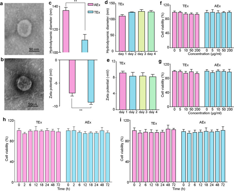
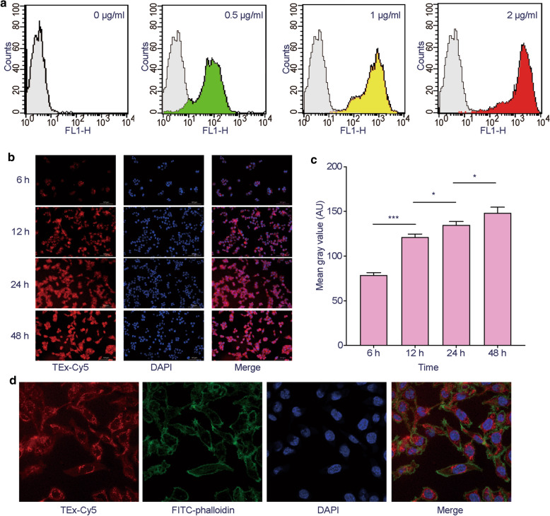
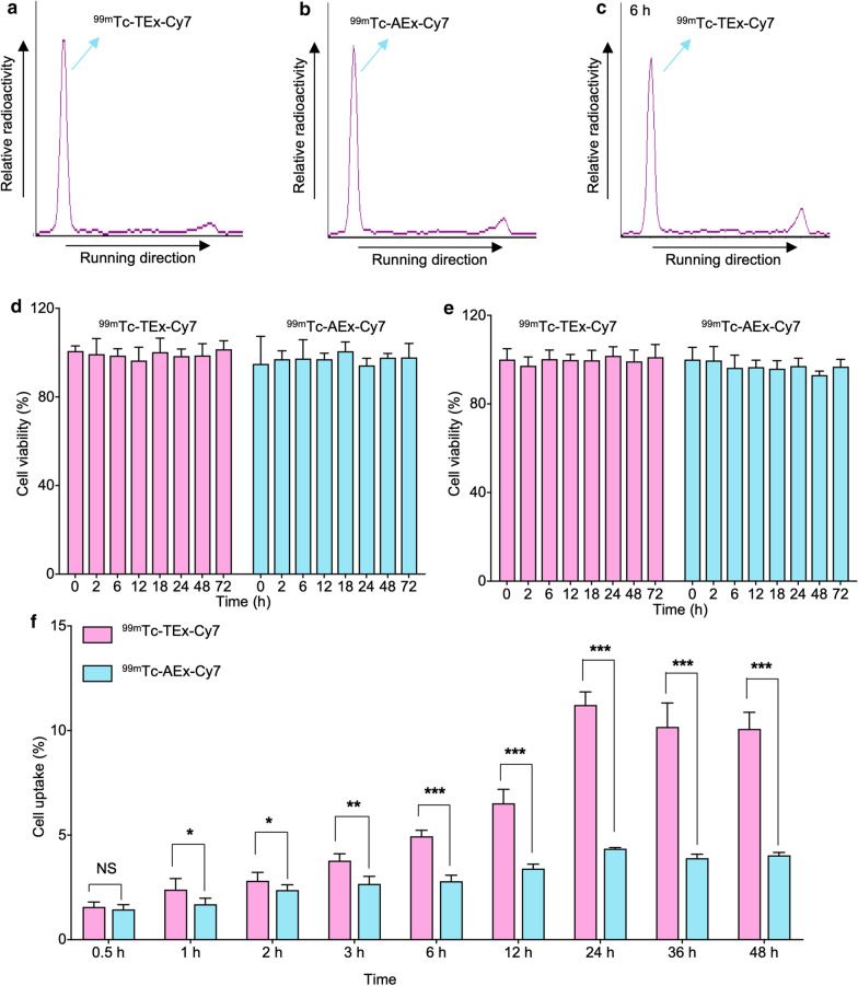
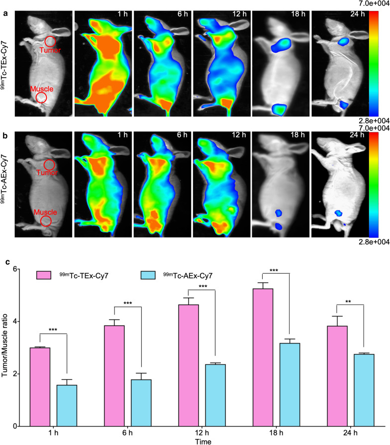
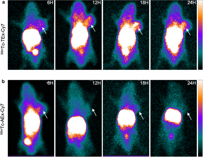
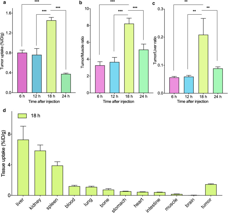
TEx were successfully isolated from HCT116 supernatants, and their membrane vesicle structure was confirmed by TEM. The average hydrodynamic diameter and zeta potential of TEx were 110.87 ± 4.61 nm and -9.20 ± 0.41 mV, respectively. Confocal microscopy and flow cytometry findings confirmed the high tumor binding ability of TEx. The uptake rate of Tc-TEx-Cy7 by HCT116 cells increased over time, reaching 14.07 ± 1.31% at 6 h of co-incubation. NIRF and SPECT imaging indicated that the most appropriate imaging time was 18 h after the injection of Tc-TEx-Cy7 when the tumor uptake (1.46% ± 0.06% ID/g) and tumor-to-muscle ratio (8.22 ± 0.65) peaked. Compared with radiolabeled adipose stem cell derived exosomes (Tc-AEx-Cy7), Tc-TEx-Cy7 exhibited a significantly higher tumor accumulation in tumor-bearing mice.
Hydrophobic insertion-based engineering of TEx may represent a promising approach to develop and label exosomes for use as nanoprobes in dual SPECT/NIRF imaging. Our findings confirmed that TEx has a higher tumor-targeting ability than AEx and highlight the potential usefulness of exosomes in biomedical applications.
肿瘤细胞衍生的外泌体(TEx)已成为有前途的药物递送纳米载体。通过非侵入性多模态成像来追踪 TEx 的体内转运可能会加速它们的临床转化。在这项研究中,我们通过疏水插入机制开发了一种基于 TEx 的纳米探针,并评估了其在结直肠癌的双单光子发射计算机断层扫描(SPECT)和近红外荧光(NIRF)成像中的性能。
TEx 从 HCT116 上清液中成功分离出来,并通过 TEM 确认了其膜囊泡结构。TEx 的平均水动力直径和 Zeta 电位分别为 110.87±4.61nm 和-9.20±0.41mV。共聚焦显微镜和流式细胞术的结果证实了 TEx 对肿瘤的高结合能力。Tc-TEx-Cy7 被 HCT116 细胞的摄取率随时间增加,在共孵育 6 小时时达到 14.07±1.31%。NIRF 和 SPECT 成像表明,在注射 Tc-TEx-Cy7 后 18 小时是最佳的成像时间,此时肿瘤摄取量(1.46%±0.06%ID/g)和肿瘤与肌肉的比值(8.22±0.65)达到峰值。与放射性标记的脂肪干细胞衍生的外泌体(Tc-AEx-Cy7)相比,Tc-TEx-Cy7 在荷瘤小鼠中表现出更高的肿瘤蓄积。
基于疏水插入的 TEx 工程可能是开发和标记外泌体作为双 SPECT/NIRF 成像中纳米探针的一种有前途的方法。我们的研究结果证实,TEx 比 AEx 具有更高的肿瘤靶向能力,并强调了外泌体在生物医学应用中的潜在用途。