Department of Nuclear Medicine, Union Hospital, Tongji Medical College, Huazhong University of Science and Technology, No. 1277 Jiefang Ave, Wuhan, 430022, Hubei Province, China.
Department of Pancreatic Surgery, Union Hospital, Tongji Medical College, Huazhong University of Science and Technology, Wuhan, 430022, China.
J Nanobiotechnology. 2021 May 22;19(1):151. doi: 10.1186/s12951-021-00888-3.
Colon cancer contributes to high mortality rates as the result of incomplete resection in tumor surgery. Multimodal imaging can provide preoperative evaluation and intraoperative image-guiding. As biocompatible nanocarriers, extracellular vesicles hold great promise for multimodal imaging. In this study, we aim to synthesized an extracellular vesicles-based nanoprobe to visualize colon cancer with positron-emission tomography/computed tomography (PET/CT) and near-infrared fluorescence (NIRF) imaging, and investigated its utility in image-guided surgery of colon cancer in animal models.
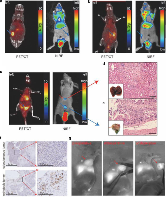
Extracellular vesicles were successfully isolated from adipose-derived stem cells (ADSCs), and their membrane vesicles were observed under TEM. DLS detected that the hydrodynamic diameters of the extracellular vesicles were approximately 140 nm and the zeta potential was - 7.93 ± 0.24 mV. Confocal microscopy showed that extracellular vesicles had a strong binding ability to tumor cells. A click chemistry-based pre-targeting strategy was used to achieve PET imaging in vivo. PET images and the biodistribution results showed that the best pre-targeting time was 20 h, and the best imaging time was 2 h after the injection of Ga-L-NETA-DBCO. The NIRF images showed that the tumor had clear images at all time points after administration of nanoparticles and the Tumor/Muscle ratio peaked at 20 h after injection. Our data also showed that both PET/CT and NIRF imaging clearly visualized the orthotopic colon cancer models, providing preoperative evaluation. Under real-time NIRF imaging, the tumor location and tumor boundary could be clearly observed.
In brief, this novel nanoprobe may be useful for multi-modal imaging of colon cancer and NIRF image-guided surgery. More importantly, this study provides a new possibility for clinical application of extracellular vesicles as nanocarriers.
结肠癌由于肿瘤手术中切除不完全导致死亡率高。多模态成像可以提供术前评估和术中图像引导。作为生物相容性纳米载体,细胞外囊泡在多模态成像中具有很大的应用前景。在本研究中,我们旨在合成一种基于细胞外囊泡的纳米探针,用于对结肠癌进行正电子发射断层扫描/计算机断层扫描(PET/CT)和近红外荧光(NIRF)成像,并研究其在动物模型中对结肠癌的图像引导手术中的应用。
成功地从脂肪来源干细胞(ADSCs)中分离出细胞外囊泡,并在 TEM 下观察到其膜囊泡。DLS 检测到细胞外囊泡的水动力直径约为 140nm,zeta 电位为-7.93±0.24mV。共聚焦显微镜显示细胞外囊泡对肿瘤细胞具有很强的结合能力。采用点击化学的前靶向策略实现体内 PET 成像。PET 图像和生物分布结果表明,最佳的前靶向时间为 20h,注射[68Ga]Ga-L-NETA-DBCO 后最佳的成像时间为 2h。NIRF 图像显示,在给予纳米颗粒后,所有时间点肿瘤均有清晰的图像,注射后 20h 肿瘤/肌肉比值达到峰值。我们的数据还表明,PET/CT 和 NIRF 成像均能清晰地显示原位结肠癌模型,提供术前评估。在实时 NIRF 成像下,可以清楚地观察到肿瘤位置和肿瘤边界。
总之,这种新型纳米探针可能对结肠癌的多模态成像和 NIRF 图像引导手术有用。更重要的是,本研究为细胞外囊泡作为纳米载体的临床应用提供了新的可能性。