Department of Medical Imaging Center, State Key Laboratory of Oncology in South China, Collaborative Innovation Center for Cancer Medicine, Sun Yat-sen University Cancer Center, 651 Dongfeng Road East, Guangzhou, Guangdong, 510060, People's Republic of China.
Department of Radiology, The University of Hong Kong-Shenzhen Hospital, No.1, Haiyuan Road Futian District, Shenzhen, 518000, People's Republic of China.
Cancer Imaging. 2021 Jan 6;21(1):2. doi: 10.1186/s40644-020-00375-2.
To determine the predictive CT imaging features for diagnosis in patients with primary pulmonary mucoepidermoid carcinomas (PMECs).
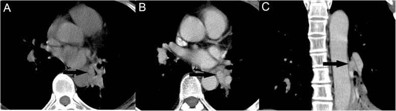
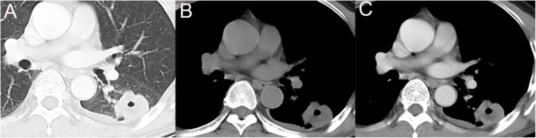
CT imaging features of 37 patients with primary PMECs, 76 with squamous cell carcinomas (SCCs) and 78 with adenocarcinomas were retrospectively reviewed. The difference of CT features among the PMECs, SCCs and adenocarcinomas was analyzed using univariate analysis, followed by multinomial logistic regression and receiver operating characteristic (ROC) curve analysis.
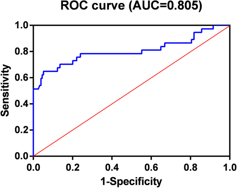
CT imaging features including tumor size, location, margin, shape, necrosis and degree of enhancement were significant different among the PMECs, SCCs and adenocarcinomas, as determined by univariate analysis (P < 0.05). Only lesion location, shape, margin and degree of enhancement remained independent factors in multinomial logistic regression analysis. ROC curve analysis showed that the area under curve of the obtained multinomial logistic regression model was 0.805 (95%CI: 0.704-0.906).
The prediction model derived from location, margin, shape and degree of enhancement can be used for preoperative diagnosis of PMECs.
旨在确定用于原发性肺黏液表皮样癌(PMEC)患者诊断的预测性 CT 成像特征。
回顾性分析了 37 例原发性 PMEC、76 例鳞状细胞癌(SCC)和 78 例腺癌患者的 CT 成像特征。采用单因素分析比较 PMEC、SCC 和腺癌之间 CT 特征的差异,然后进行多项逻辑回归和受试者工作特征(ROC)曲线分析。
通过单因素分析(P<0.05),发现 CT 成像特征包括肿瘤大小、位置、边缘、形状、坏死和强化程度在 PMEC、SCC 和腺癌之间存在显著差异。多项逻辑回归分析显示,病变位置、形状、边缘和强化程度仍然是独立因素。ROC 曲线分析表明,该多项逻辑回归模型的曲线下面积为 0.805(95%CI:0.704-0.906)。
源自位置、边缘、形状和强化程度的预测模型可用于 PMEC 的术前诊断。