Ribas Beatriz Ribeiro, Nascimento Eduarda Helena Leandro, Freitas Deborah Queiroz, Pontual Andréa Dos Anjos, Pontual Maria Luiza Dos Anjos, Perez Danyel Elias Cruz, Ramos-Perez Flávia Maria Moraes
Department of Clinical and Preventive Dentistry, Federal University of Pernambuco, Recife, Pernambuco, Brazil.
Division of Oral Radiology, Department of Oral Diagnosis, Piracicaba Dental School, University of Campinas, Piracicaba, Sao Paulo, Brazil.
Imaging Sci Dent. 2020 Dec;50(4):281-290. doi: 10.5624/isd.2020.50.4.281. Epub 2020 Dec 15.
The objective of the present study was to evaluate the prevalence of dental implants positioning errors and their associations with adjacent structures and anatomical variations by means of cone-beam computed tomography (CBCT).
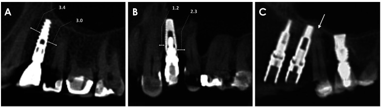
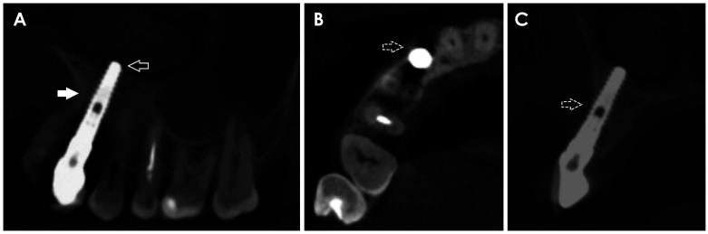
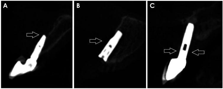
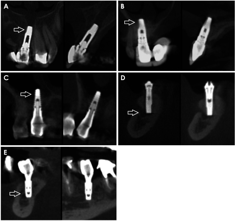
CBCT images of 207 patients (584 dental implants) were evaluated by 2 oral radiologists. The distance between the implant and the adjacent teeth/implants was measured and classified as adequate (≥1.5 mm and ≥3 mm, respectively) or inadequate. The presence of thread exposure, cortical perforation, implant dehiscence, implant penetration into adjacent structures, and anatomical variations was also recorded. The incisor canal diameter and the depth of the concavity of the submandibular fossa were measured in order to evaluate their correlations with the frequency of implant penetration in these structures. Descriptive analyses, the Fisher exact test, and Spearman correlation analysis were performed (α=0.05).
The overall prevalence of positioning errors was 82.9%. The most common error was the inadequate distance between the implant and the adjacent teeth/implants. The presence of anatomical variations did not significantly influence the overall prevalence of errors (>0.05). There was a positive correlation between the diameter of the incisor canal and the frequency of implant penetration in this structure (r=0.232, <0.05).
There was a high prevalence of dental implant positioning errors, and positioning errors were not associated with the presence of anatomical variations. Professionals should be aware of the space available for implant placement during the preoperative planning stage.
本研究的目的是通过锥形束计算机断层扫描(CBCT)评估牙种植体定位错误的发生率及其与相邻结构和解剖变异的关联。
2名口腔放射科医生对207例患者(584颗牙种植体)的CBCT图像进行评估。测量种植体与相邻牙齿/种植体之间的距离,并将其分类为足够(分别≥1.5mm和≥3mm)或不足。还记录了螺纹暴露、皮质穿孔、种植体裂开、种植体穿入相邻结构以及解剖变异的情况。测量切牙管直径和下颌下窝凹陷深度,以评估它们与这些结构中种植体穿入频率的相关性。进行描述性分析、Fisher精确检验和Spearman相关性分析(α=0.05)。
定位错误的总体发生率为82.9%。最常见的错误是种植体与相邻牙齿/种植体之间的距离不足。解剖变异的存在并未显著影响错误的总体发生率(>0.05)。切牙管直径与该结构中种植体穿入频率之间存在正相关(r=0.232,<0.05)。
牙种植体定位错误的发生率很高,且定位错误与解剖变异的存在无关。专业人员在术前规划阶段应了解种植体植入的可用空间。