Lindner Melanie, Verhagen Irene, Viitaniemi Heidi M, Laine Veronika N, Visser Marcel E, Husby Arild, van Oers Kees
Department of Animal Ecology, Netherlands Institute of Ecology (NIOO-KNAW), P.O. Box 50, Wageningen, 6700, AB, The Netherlands.
Chronobiology Unit, Groningen Institute for Evolutionary Life Sciences (GELIFES), University of Groningen, Groningen, The Netherlands.
BMC Genomics. 2021 Jan 7;22(1):36. doi: 10.1186/s12864-020-07329-9.
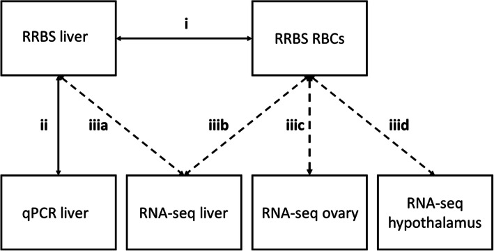
DNA methylation is likely a key mechanism regulating changes in gene transcription in traits that show temporal fluctuations in response to environmental conditions. To understand the transcriptional role of DNA methylation we need simultaneous within-individual assessment of methylation changes and gene expression changes over time. Within-individual repeated sampling of tissues, which are essential for trait expression is, however, unfeasible (e.g. specific brain regions, liver and ovary for reproductive timing). Here, we explore to what extend between-individual changes in DNA methylation in a tissue accessible for repeated sampling (red blood cells (RBCs)) reflect such patterns in a tissue unavailable for repeated sampling (liver) and how these DNA methylation patterns are associated with gene expression in such inaccessible tissues (hypothalamus, ovary and liver). For this, 18 great tit (Parus major) females were sacrificed at three time points (n = 6 per time point) throughout the pre-laying and egg-laying period and their blood, hypothalamus, ovary and liver were sampled.
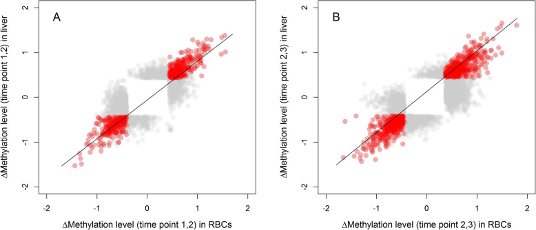
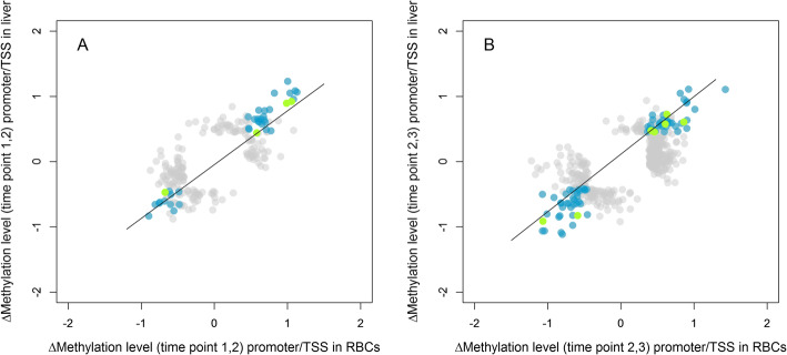
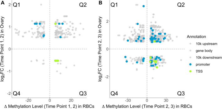
We simultaneously assessed DNA methylation changes (via reduced representation bisulfite sequencing) and changes in gene expression (via RNA-seq and qPCR) over time. In general, we found a positive correlation between changes in CpG site methylation in RBCs and liver across timepoints. For CpG sites in close proximity to the transcription start site, an increase in RBC methylation over time was associated with a decrease in the expression of the associated gene in the ovary. In contrast, no such association with gene expression was found for CpG site methylation within the gene body or the 10 kb up- and downstream regions adjacent to the gene body.
Temporal changes in DNA methylation are largely tissue-general, indicating that changes in RBC methylation can reflect changes in DNA methylation in other, often less accessible, tissues such as the liver in our case. However, associations between temporal changes in DNA methylation with changes in gene expression are mostly tissue- and genomic location-dependent. The observation that temporal changes in DNA methylation within RBCs can relate to changes in gene expression in less accessible tissues is important for a better understanding of how environmental conditions shape traits that temporally change in expression in wild populations.
DNA甲基化可能是一种关键机制,用于调节那些因环境条件而呈现时间波动的性状中基因转录的变化。为了理解DNA甲基化的转录作用,我们需要在个体内部同时评估甲基化变化和基因表达随时间的变化。然而,对性状表达至关重要的组织进行个体内部重复采样是不可行的(例如特定脑区、用于生殖时间调控的肝脏和卵巢)。在这里,我们探讨在可重复采样的组织(红细胞(RBC))中个体间DNA甲基化变化在多大程度上反映了不可重复采样的组织(肝脏)中的此类模式,以及这些DNA甲基化模式如何与此类难以获取的组织(下丘脑、卵巢和肝脏)中的基因表达相关联。为此,在整个产卵前期和产卵期的三个时间点处死了18只雌性大山雀(Parus major)(每个时间点n = 6只),并采集了它们的血液、下丘脑、卵巢和肝脏样本。
我们同时评估了随时间变化的DNA甲基化变化(通过简化代表性亚硫酸氢盐测序)和基因表达变化(通过RNA测序和定量PCR)。总体而言,我们发现红细胞和肝脏中CpG位点甲基化随时间的变化呈正相关。对于靠近转录起始位点的CpG位点,红细胞甲基化随时间增加与卵巢中相关基因的表达减少相关。相比之下,在基因体内或与基因体相邻的10 kb上下游区域内的CpG位点甲基化与基因表达之间未发现此类关联。
DNA甲基化的时间变化在很大程度上具有组织普遍性,这表明红细胞甲基化的变化可以反映其他通常难以获取的组织(如我们研究中的肝脏)中DNA甲基化的变化。然而,DNA甲基化时间变化与基因表达变化之间的关联大多取决于组织和基因组位置。红细胞内DNA甲基化的时间变化与难以获取的组织中的基因表达变化相关这一观察结果,对于更好地理解环境条件如何塑造野生种群中表达随时间变化的性状非常重要。