Al-Bachari Sarah, Naish Josephine H, Parker Geoff J M, Emsley Hedley C A, Parkes Laura M
Lancaster Medical School, Faculty of Health and Medicine, Lancaster University, Lancaster, United Kingdom.
Department of Neurology, Lancashire Teaching Hospitals NHS Foundation Trust, Preston, United Kingdom.
Front Physiol. 2020 Dec 22;11:593026. doi: 10.3389/fphys.2020.593026. eCollection 2020.
Blood-brain barrier (BBB) disruption has been noted in animal models of Parkinson's disease (PD) and forms the basis of the vascular hypothesis of neurodegeneration, yet clinical studies are lacking.
To determine alterations in BBB integrity in PD, with comparison to cerebrovascular disease.
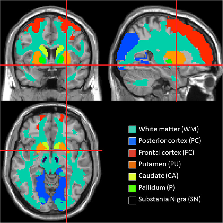
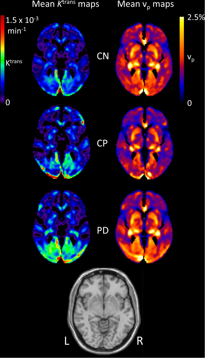
Dynamic contrast enhanced magnetic resonance images were collected from 49 PD patients, 15 control subjects with cerebrovascular disease [control positive (CP)] and 31 healthy control subjects [control negative (CN)], with all groups matched for age. Quantitative maps of the contrast agent transfer coefficient across the BBB ( ) and plasma volume (v ) were produced using Patlak analysis. Differences in and v were assessed with voxel-based analysis as well as in regions associated with PD pathophysiology. In addition, the volume of white matter lesions (WMLs) was obtained from T-weighted fluid attenuation inversion recovery (FLAIR) images.
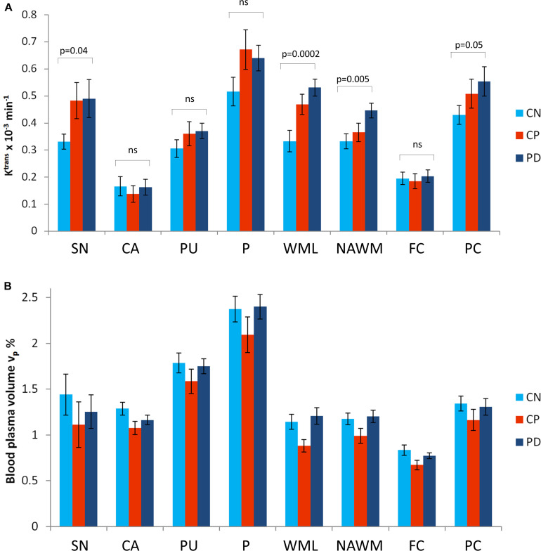
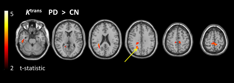
Higher , reflecting higher BBB leakage, was found in the PD group than in the CN group using voxel-based analysis; differences were most prominent in the posterior white matter regions. Region of interest analysis confirmed to be significantly higher in PD than in CN, predominantly driven by differences in the substantia nigra, normal-appearing white matter, WML and the posterior cortex. WML volume was significantly higher in PD compared to CN. values and WML volume were similar in PD and CP, suggesting a similar burden of cerebrovascular disease despite lower cardiovascular risk factors.
These results show BBB disruption in PD.
在帕金森病(PD)动物模型中已发现血脑屏障(BBB)破坏,这构成了神经退行性变血管假说的基础,但缺乏临床研究。
确定PD患者血脑屏障完整性的改变,并与脑血管疾病进行比较。
收集了49例PD患者、15例患有脑血管疾病的对照受试者[对照阳性(CP)]和31例健康对照受试者[对照阴性(CN)]的动态对比增强磁共振图像,所有组年龄匹配。使用Patlak分析生成跨血脑屏障的造影剂转移系数( )和血浆容积(v )的定量图。通过基于体素的分析以及与PD病理生理学相关的区域评估 和v 的差异。此外,从T加权液体衰减反转恢复(FLAIR)图像中获得白质病变(WMLs)的体积。
基于体素的分析发现,PD组的 高于CN组,反映出血脑屏障渗漏更高;差异在后部白质区域最为突出。感兴趣区域分析证实,PD组的 显著高于CN组,主要由黑质、外观正常的白质、WML和后部皮质的差异驱动。与CN相比,PD组的WML体积显著更高。PD组和CP组的 值和WML体积相似,表明尽管心血管危险因素较低,但脑血管疾病负担相似。
这些结果表明PD存在血脑屏障破坏。