• 文献检索
  • 文档翻译
  • 深度研究
  • 学术资讯
  • Suppr Zotero 插件Zotero 插件
  • 邀请有礼
  • 套餐&价格
  • 历史记录
应用&插件
Suppr Zotero 插件Zotero 插件浏览器插件Mac 客户端Windows 客户端微信小程序
定价
高级版会员购买积分包购买API积分包
服务
文献检索文档翻译深度研究API 文档MCP 服务
关于我们
关于 Suppr公司介绍联系我们用户协议隐私条款
关注我们

Suppr 超能文献

核心技术专利:CN118964589B侵权必究
粤ICP备2023148730 号-1Suppr @ 2026

文献检索

告别复杂PubMed语法,用中文像聊天一样搜索,搜遍4000万医学文献。AI智能推荐,让科研检索更轻松。

立即免费搜索

文件翻译

保留排版,准确专业,支持PDF/Word/PPT等文件格式,支持 12+语言互译。

免费翻译文档

深度研究

AI帮你快速写综述,25分钟生成高质量综述,智能提取关键信息,辅助科研写作。

立即免费体验

COVID-19:一种定性的胸部 CT 模型,用于识别疾病的严重形式。

COVID-19: A qualitative chest CT model to identify severe form of the disease.

机构信息

Department of Radiology, Reims University Hospital, 51092 Reims, France.

Clinical Research Department, Reims University Hospital, 51092 Reims, France.

出版信息

Diagn Interv Imaging. 2021 Feb;102(2):77-84. doi: 10.1016/j.diii.2020.12.002. Epub 2020 Dec 17.

DOI:10.1016/j.diii.2020.12.002
PMID:33419693
原文链接:https://pmc.ncbi.nlm.nih.gov/articles/PMC7746121/
Abstract

PURPOSE

The purpose of this study was to identify clinical and chest computed tomography (CT) features associated with a severe form of coronavirus disease 2019 (COVID-19) and to propose a quick and easy to use model to identify patients at risk of a severe form.

MATERIALS AND METHODS

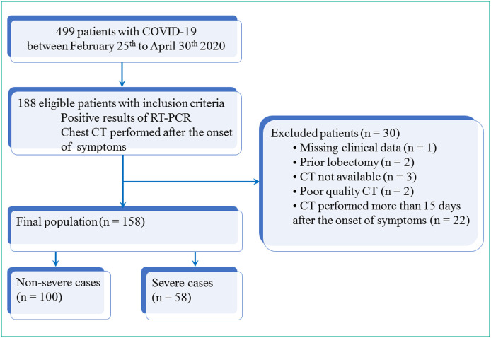
A total of 158 patients with biologically confirmed COVID-19 who underwent a chest CT after the onset of the symptoms were included. There were 84 men and 74 women with a mean age of 68±14 (SD) years (range: 24-96years). There were 100 non-severe and 58 severe cases. Their clinical data were recorded and the first chest CT examination was reviewed using a computerized standardized report. Univariate and multivariate analyses were performed in order to identify the risk factors associated with disease severity. Two models were built: one was based only on qualitative CT features and the other one included a semi-quantitative total CT score to replace the variable representing the extent of the disease. Areas under the ROC curves (AUC) of the two models were compared with DeLong's method.

RESULTS

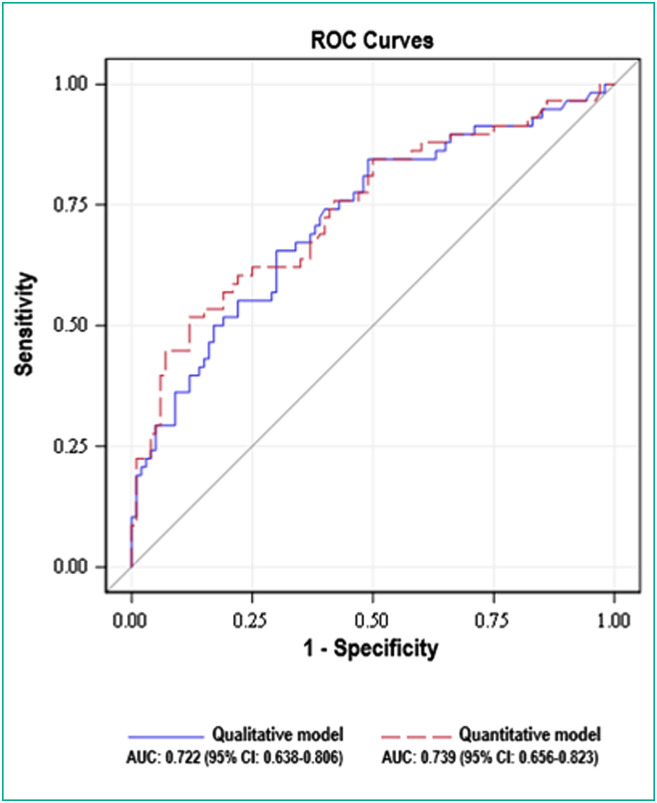
Central involvement of lung parenchyma (P<0.001), area of consolidation (P<0.008), air bronchogram sign (P<0.001), bronchiectasis (P<0.001), traction bronchiectasis (P<0.011), pleural effusion (P<0.026), large involvement of either one of the upper lobes or of the middle lobe (P<0.001) and total CT score≥15 (P<0.001) were more often observed in the severe group than in the non-severe group. No significant differences were found between the qualitative model (large involvement of either upper lobes or middle lobe [odd ratio (OR)=2.473], central involvement [OR=2.760], pleural effusion [OR=2.699]) and the semi-quantitative model (total CT score≥15 [OR=3.342], central involvement [OR=2.344], pleural effusion [OR=2.754]) with AUC of 0.722 (95% CI: 0.638-0.806) vs. 0.739 (95% CI: 0.656-0.823), respectively (P=0.209).

CONCLUSION

We have developed a new qualitative chest CT-based multivariate model that provides independent risk factors associated with severe form of COVID-19.

摘要

目的

本研究旨在确定与 2019 年冠状病毒病(COVID-19)严重形式相关的临床和胸部计算机断层扫描(CT)特征,并提出一种快速易用的模型来识别有严重形式风险的患者。

材料和方法

共纳入 158 例经生物学证实的 COVID-19 患者,这些患者在症状出现后均接受了胸部 CT 检查。其中男性 84 例,女性 74 例,平均年龄 68±14(SD)岁(范围:24-96 岁)。100 例为非重症病例,58 例为重症病例。记录了他们的临床数据,并使用计算机化的标准化报告对首次胸部 CT 检查进行了回顾。进行了单变量和多变量分析,以确定与疾病严重程度相关的危险因素。建立了两个模型:一个仅基于定性 CT 特征,另一个模型则包含半定量总 CT 评分来代替代表疾病程度的变量。使用 DeLong 方法比较了两个模型的 ROC 曲线下面积(AUC)。

结果

肺部实质中央受累(P<0.001)、实变区面积(P<0.008)、空气支气管征(P<0.001)、支气管扩张(P<0.001)、牵拉性支气管扩张(P<0.011)、胸腔积液(P<0.026)、单侧上叶或中叶的大受累(P<0.001)和总 CT 评分≥15(P<0.001)在重症组中比在非重症组中更常见。定性模型(单侧上叶或中叶大受累[比值比(OR)=2.473]、中央受累[OR=2.760]、胸腔积液[OR=2.699])和半定量模型(总 CT 评分≥15[OR=3.342]、中央受累[OR=2.344]、胸腔积液[OR=2.754])与 AUC 为 0.722(95%CI:0.638-0.806)和 0.739(95%CI:0.656-0.823),分别为 0.722(95%CI:0.638-0.806)和 0.739(95%CI:0.656-0.823)(P=0.209),之间无显著差异。

结论

我们开发了一种新的基于胸部 CT 的多变量定性模型,提供了与 COVID-19 严重形式相关的独立危险因素。

https://cdn.ncbi.nlm.nih.gov/pmc/blobs/4118/7746121/28c0018cecc9/gr4_lrg.jpg
https://cdn.ncbi.nlm.nih.gov/pmc/blobs/4118/7746121/ca23a14ad7f0/gr1_lrg.jpg
https://cdn.ncbi.nlm.nih.gov/pmc/blobs/4118/7746121/77284fe340b3/gr2_lrg.jpg
https://cdn.ncbi.nlm.nih.gov/pmc/blobs/4118/7746121/a87df16dcd30/gr3_lrg.jpg
https://cdn.ncbi.nlm.nih.gov/pmc/blobs/4118/7746121/28c0018cecc9/gr4_lrg.jpg

相似文献

1
COVID-19: A qualitative chest CT model to identify severe form of the disease.COVID-19:一种定性的胸部 CT 模型,用于识别疾病的严重形式。
Diagn Interv Imaging. 2021 Feb;102(2):77-84. doi: 10.1016/j.diii.2020.12.002. Epub 2020 Dec 17.
2
The Performance of Chest CT in Evaluating the Clinical Severity of COVID-19 Pneumonia: Identifying Critical Cases Based on CT Characteristics.胸部 CT 对评估 COVID-19 肺炎临床严重程度的性能:基于 CT 特征识别重症病例。
Invest Radiol. 2020 Jul;55(7):412-421. doi: 10.1097/RLI.0000000000000689.
3
Analysis of clinical features and imaging signs of COVID-19 with the assistance of artificial intelligence.人工智能辅助分析 COVID-19 的临床特征和影像征象。
Eur Rev Med Pharmacol Sci. 2020 Aug;24(15):8210-8218. doi: 10.26355/eurrev_202008_22510.
4
A new imaging sign in COVID-19 pneumonia: vascular changes and their correlation with clinical severity of the disease.新型冠状病毒肺炎的一种新影像学征象:血管改变及其与疾病严重程度的相关性。
Diagn Interv Radiol. 2021 Mar;27(2):172-180. doi: 10.5152/dir.2020.20346.
5
CT Features of Coronavirus Disease 2019 (COVID-19) Pneumonia in 62 Patients in Wuhan, China.中国武汉 62 例 2019 年冠状病毒病(COVID-19)肺炎的 CT 特征。
AJR Am J Roentgenol. 2020 Jun;214(6):1287-1294. doi: 10.2214/AJR.20.22975. Epub 2020 Mar 5.
6
A Comparison of Clinical and Chest CT Findings in Patients With Influenza A (H1N1) Virus Infection and Coronavirus Disease (COVID-19).甲型流感病毒(H1N1)感染与新型冠状病毒肺炎(COVID-19)患者的临床和胸部 CT 表现比较。
AJR Am J Roentgenol. 2020 Nov;215(5):1065-1071. doi: 10.2214/AJR.20.23214. Epub 2020 May 26.
7
Six-month Follow-up Chest CT Findings after Severe COVID-19 Pneumonia.重症 COVID-19 肺炎患者 6 个月随访的胸部 CT 结果。
Radiology. 2021 Apr;299(1):E177-E186. doi: 10.1148/radiol.2021203153. Epub 2021 Jan 26.
8
Prognostic Significance of Chest Imaging by LUS and CT in COVID-19 Inpatients: The ECOVID Multicenter Study.超声与 CT 肺部影像学对 COVID-19 住院患者预后评估的意义:ECCOVID 多中心研究。
Respiration. 2022;101(2):122-131. doi: 10.1159/000518516. Epub 2021 Aug 31.
9
Chest CT in the Emergency Department for Diagnosis of COVID-19 Pneumonia: Dutch Experience.急诊科胸部 CT 诊断 COVID-19 肺炎:荷兰经验。
Radiology. 2021 Feb;298(2):E98-E106. doi: 10.1148/radiol.2020203465. Epub 2020 Nov 17.
10
Value of quantitative analysis in lung computed tomography in patients severely ill with COVID-19.COVID-19 重症患者肺部计算机断层扫描的定量分析价值。
PLoS One. 2021 May 20;16(5):e0251946. doi: 10.1371/journal.pone.0251946. eCollection 2021.

引用本文的文献

1
Quantitative Chest CT Analysis: Three Different Approaches to Quantify the Burden of Viral Interstitial Pneumonia Using COVID-19 as a Paradigm.胸部CT定量分析:以COVID-19为范例的三种不同方法量化病毒性间质性肺炎的负担
J Clin Med. 2024 Dec 1;13(23):7308. doi: 10.3390/jcm13237308.
2
Time-dependent CT score-based model for identifying severe/critical COVID-19 at a fever clinic after the emergence of Omicron variant.基于时间依赖性CT评分的模型,用于在奥密克戎变异株出现后在发热门诊识别重症/危重症新冠肺炎
Heliyon. 2024 Mar 25;10(7):e27963. doi: 10.1016/j.heliyon.2024.e27963. eCollection 2024 Apr 15.
3
Computed tomography severity score as a predictor of disease severity and mortality in COVID-19 patients: A systematic review and meta-analysis.
计算机断层扫描严重程度评分作为 COVID-19 患者疾病严重程度和死亡率的预测指标:系统评价和荟萃分析。
J Med Imaging Radiat Sci. 2023 Jun;54(2):364-375. doi: 10.1016/j.jmir.2023.02.003. Epub 2023 Feb 16.
4
Short and Long-Term Impact of COVID-19 Infection on Previous Respiratory Diseases.COVID-19 感染对既往呼吸道疾病的短期和长期影响。
Arch Bronconeumol. 2022 Apr;58 Suppl 1:39-50. doi: 10.1016/j.arbres.2022.03.011. Epub 2022 Apr 15.
5
Triaging of COVID-19 patients using low dose chest CT: Incidence and factor analysis of lung involvement on CT images.采用低剂量胸部 CT 对 COVID-19 患者进行分诊:CT 图像上肺部受累的发生率及相关因素分析。
J Infect Chemother. 2022 Jun;28(6):797-801. doi: 10.1016/j.jiac.2022.02.025. Epub 2022 Mar 14.
6
The Multifaceted COVID-19: CT Aspects of Its Atypical Pulmonary and Abdominal Manifestations and Complications in Adults and Children. A Pictorial Review.多面的 COVID-19:成人和儿童非典型肺部及腹部表现和并发症的 CT 特征。图文综述。
Microorganisms. 2021 Sep 26;9(10):2037. doi: 10.3390/microorganisms9102037.
7
Imaging of COVID-19: An update of current evidences.COVID-19 的影像学表现:现有证据的更新。
Diagn Interv Imaging. 2021 Sep;102(9):493-500. doi: 10.1016/j.diii.2021.05.006. Epub 2021 May 25.
8
COVID-19 pneumonia: The fight must go on.新冠肺炎:战斗必须继续。
Diagn Interv Imaging. 2021 Feb;102(2):61-62. doi: 10.1016/j.diii.2021.01.006.