Shash Hani Ali, Almarzouq Sawsan Fahad, Alghamdi Abdulrahman Abdulaziz, Alratroot Jumana Abdulwahab
Department of Surgery, Plastic Surgery, College of Medicine, Imam Abdulrahman Bin Faisal University, Dammam, Saudi Arabia.
College of Medicine, Imam Abdulrahman Bin Faisal University, Dammam, Saudi Arabia.
Plast Reconstr Surg Glob Open. 2020 Dec 3;8(12):e3234. doi: 10.1097/GOX.0000000000003234. eCollection 2020 Dec.
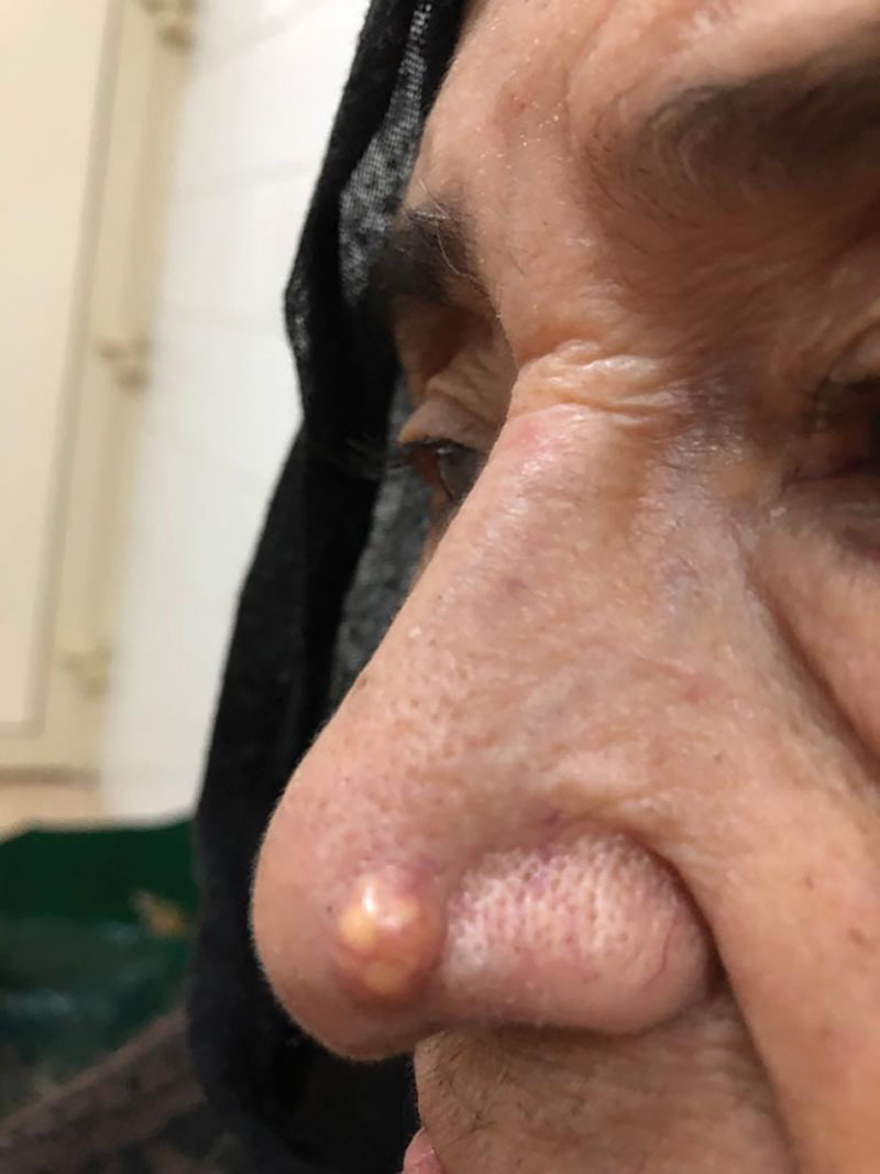
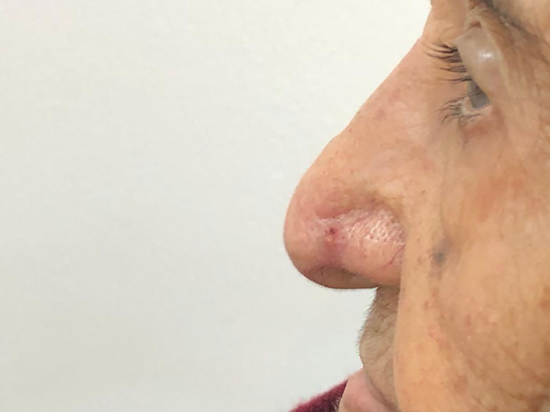
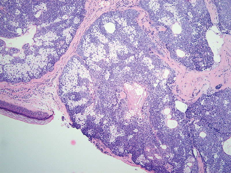
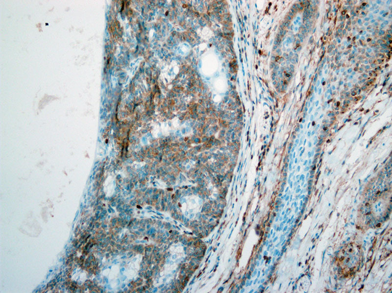
We report a very rare type of tumor in the left nasal ala in an elderly patient. An 81-year-old Saudi woman known to have hypertension, osteoporosis, and rheumatoid disease (who had been compliant to her medications) presented with a 0.5-cm fixed, firm, round well-defined nodule on the left ala of the nose (with crusting, erosion, and telangiectasia of the overlying skin), whose size had been gradually increasing for 2 years. The patient underwent excisional biopsy, and the specimen was sent for a histopathologic analysis. Macroscopic examination showed a round tan-white homogenous nodule, measuring 0.6 × 0.5 × 0.5 cm. Microscopic examination revealed a fairly circumscribed unencapsulated dermal lesion, featuring basaloid cells with peripheral palisading, and focal stromal clefting. The final diagnosis of basal cell carcinoma with sebaceous differentiation was made. The patient was managed with Mohs surgery with clear margins, and full-thickness skin graft was done. Four months after surgery, the patient had a recurrence, which was managed with a surgical excision (with 4-mm margin) and covered by a full-thickness skin graft.
我们报告了一例老年患者左侧鼻翼出现的一种非常罕见的肿瘤。一名81岁的沙特女性,患有高血压、骨质疏松症和类风湿疾病(一直遵医嘱服药),其左侧鼻翼出现一个0.5厘米大小、固定、质地硬、圆形且边界清晰的结节(其上覆皮肤有结痂、糜烂和毛细血管扩张),该结节大小已逐渐增大两年。患者接受了切除活检,标本送去做组织病理学分析。肉眼检查显示为一个圆形的棕白色均匀结节,大小为0.6×0.5×0.5厘米。显微镜检查发现是一个边界相当清晰的无包膜真皮病变,特征为基底样细胞呈周边栅栏状排列,并有局灶性间质裂隙。最终诊断为伴有皮脂腺分化的基底细胞癌。患者接受了切缘清晰的莫氏手术,并进行了全厚皮片移植。术后四个月,患者复发,再次接受手术切除(切缘4毫米),并用全厚皮片覆盖。