Iimori Yasumasa, Iwai Ryosuke, Nagatani Kengo, Inoue Yuka, Funayama-Iwai Marina, Okamoto Mari, Nakata Mio, Mie Keiichiro, Nishida Hidetaka, Nakayama Yasuhide, Akiyoshi Hideo
Laboratory of Veterinary Surgery, Graduate School of Life and Environmental Sciences, Osaka Prefecture University, 1-58 Rinku-oraikita, Izumisano-shi, Osaka 598-8531, Japan.
Research Institute of Technology, Okayama University of Science, 1-1 Ridaicho, Kita-ku, Okayama, 700-0005, Japan.
Regen Ther. 2020 Nov 20;15:274-280. doi: 10.1016/j.reth.2020.10.006. eCollection 2020 Dec.
In-body tissue architecture (iBTA) technology, based on cell-free tissue engineering, can produces collagenous tissues for implantation by subcutaneous embedding a designed mold. The aim of this study was to evaluate the biocompatibility of iBTA-induced "Biosheet®" collagenous sheets, as scaffold materials for bladder reconstruction.
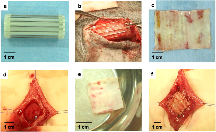
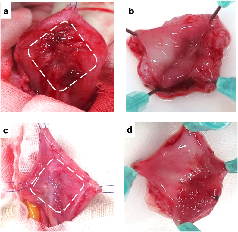
Canine Biosheet® implants were prepared by embedding molds into subcutaneous pouches in beagles for 8 weeks. A part of canine bladder wall was excised (2 × 2 cm) and repaired by patching the same sized autologous Biosheet®. The Biosheet® implants were harvested 4 weeks (n = 1) and 12 weeks (n = 3) after the implantation and evaluated histologically.
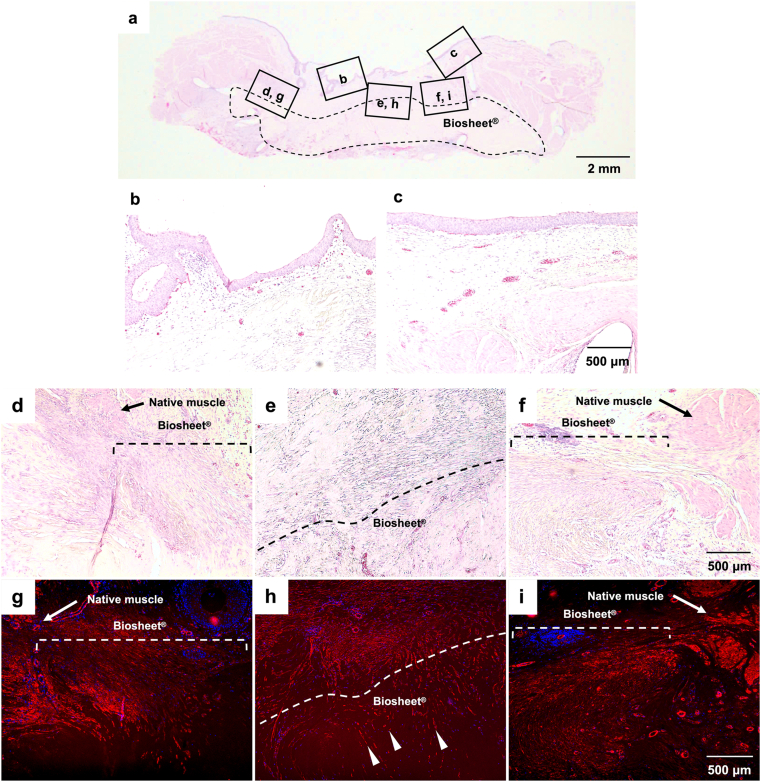
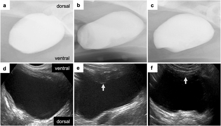
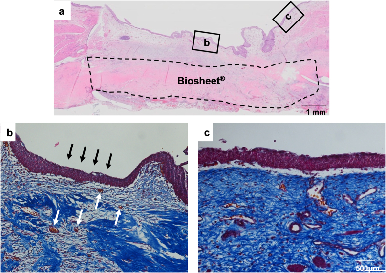
No disruption of the patched Biosheet® implants or urinary leakage into the peritoneal cavity was observed during the entire observation periods. There were no signs of chronic inflammation or Biosheet® rejection. The urine-contacting surface of luminal surface of the Biosheet® was covered with a multicellular layer of urothelium cells 4 weeks after implantation. α-SMA-positive muscle cells were observed at the margin of the Biosheet® implants at 12 weeks after the implantation. In addition, in the center of the Biosheet® implants, the formation of microvessels stained as α-SMA-positive was observed.
Biosheet® implants have biocompatibility as a scaffold for bladder reconstruction, indicating that they may be applicable for full-thickness bladder wall substitution. Further studies are required for definitive evaluation as a scaffold for bladder reconstruction.
基于无细胞组织工程的体内组织结构(iBTA)技术,能够通过皮下植入设计好的模具来生产用于植入的胶原组织。本研究的目的是评估iBTA诱导的“生物片®”胶原片作为膀胱重建支架材料的生物相容性。
通过将模具植入比格犬的皮下袋8周来制备犬生物片®植入物。切除部分犬膀胱壁(2×2厘米),并用相同大小的自体生物片®进行修补。在植入后4周(n = 1)和12周(n = 3)收集生物片®植入物,并进行组织学评估。
在整个观察期内,未观察到修补的生物片®植入物破裂或尿液漏入腹腔。没有慢性炎症或生物片®排斥的迹象。植入后4周,生物片®腔面的尿液接触表面覆盖有多层尿路上皮细胞。植入后12周,在生物片®植入物边缘观察到α-SMA阳性肌细胞。此外,在生物片®植入物中心,观察到α-SMA阳性染色的微血管形成。
生物片®植入物作为膀胱重建支架具有生物相容性,表明它们可能适用于全层膀胱壁替代。作为膀胱重建支架进行确定性评估还需要进一步研究。