Dai Dan, Lacadie Cheryl M, Holmes Sophie E, Cool Ryan, Anticevic Alan, Averill Chris, Abdallah Chadi, Esterlis Irina
Department of Psychiatry, Yale University School of Medicine, New Haven, Connecticut.
Department of Radiology and Biomedical Imaging, Yale University School of Medicine, New Haven, Connecticut.
Chronic Stress (Thousand Oaks). 2020 Dec 22;4:2470547020980681. doi: 10.1177/2470547020980681. eCollection 2020 Jan-Dec.
Ketamine is a novel fast-acting antidepressant. Acute ketamine treatment can reverse microstructure deficits and normalize functional alterations in the brain, but little is known about the impacts of ketamine on brain volumes in individuals with depression.
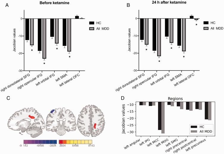
We used 3 T magnetic resonance imaging (MRI) and tensorbased morphological methods to investigate the regional volume differences for 29 healthy control (HC) subjects and 21 subjects with major depressive disorder (MDD), including 10 subjects with comorbid post-traumatic stress disorder (PTSD). All the subjects participated in MRI scanning before and 24 h post intravenous ketamine infusion. The effects of acute ketamine administration on HC, MDD, and MDD/PTSD groups were examined separately by whole-brain voxel-wise t-tests.
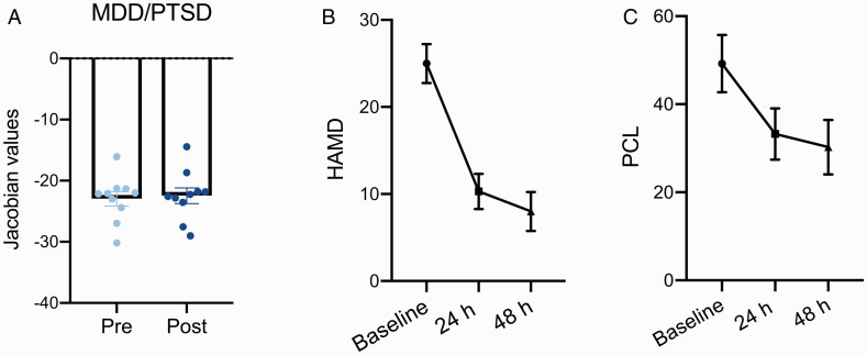
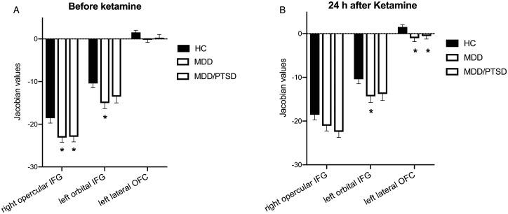
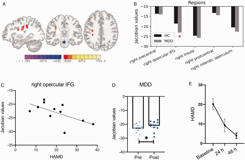
Our data showed smaller volume of inferior frontal gyrus (IFG, opercular part) in MDD and MDD/PTSD subjects compared to HC, and a significant correlation between opercular IFG volume and depressive severity in MDD subjects only. Ketamine administration normalized the structural alterations of opercular IFG in both MDD and MDD/PTSD groups, and significantly improved depressive and PTSD symptoms. Twenty-four hours after a single ketamine infusion, there were two clusters of voxels with volume changes in MDD subjects, including significantly increased volumes of opercular IFG. No significant structural alterations were found in the MDD/PTSD or HC groups.
These findings provide direct evidence that acute ketamine administration can normalize structural alterations associated with depression and highlight the importance of IFG in the guidance of future therapeutic targets.
氯胺酮是一种新型速效抗抑郁药。急性氯胺酮治疗可逆转大脑中的微观结构缺陷并使功能改变正常化,但关于氯胺酮对抑郁症患者脑容量的影响知之甚少。
我们使用3T磁共振成像(MRI)和基于张量的形态学方法,对29名健康对照(HC)受试者和21名重度抑郁症(MDD)受试者(包括10名合并创伤后应激障碍(PTSD)的受试者)的区域体积差异进行了研究。所有受试者在静脉注射氯胺酮前和注射后24小时均接受了MRI扫描。通过全脑体素水平的t检验分别检查急性氯胺酮给药对HC、MDD和MDD/PTSD组的影响。
我们的数据显示,与HC相比,MDD和MDD/PTSD受试者的额下回(IFG,岛盖部)体积较小,且仅在MDD受试者中,岛盖部IFG体积与抑郁严重程度之间存在显著相关性。氯胺酮给药使MDD和MDD/PTSD组岛盖部IFG的结构改变正常化,并显著改善了抑郁和PTSD症状。单次注射氯胺酮24小时后,MDD受试者中有两簇体素出现体积变化,包括岛盖部IFG体积显著增加。在MDD/PTSD或HC组中未发现明显的结构改变。
这些发现提供了直接证据,表明急性氯胺酮给药可使与抑郁症相关的结构改变正常化,并突出了IFG在未来治疗靶点指导中的重要性。