Blatt Sebastian, Thiem Daniel G E, Pabst Andreas, Al-Nawas Bilal, Kämmerer Peer W
Department of Oral and Maxillofacial Surgery, University Medical Center, Augustusplatz 2, 55131 Mainz, Germany.
Platform for Biomaterial Research, BiomaTiCS Group, University Medical Center, Langenbeckstrasse 1, 55131 Mainz, Germany.
Biomedicines. 2021 Jan 10;9(1):61. doi: 10.3390/biomedicines9010061.
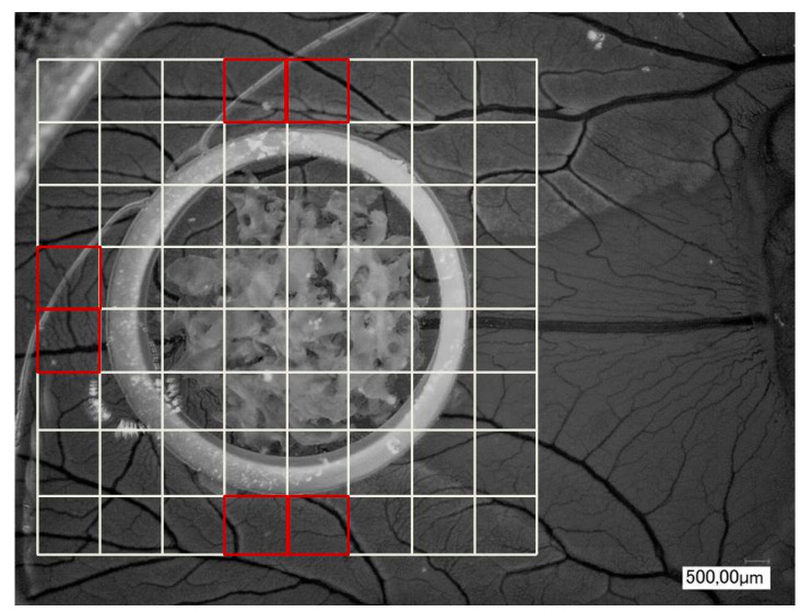
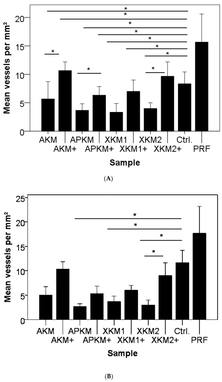
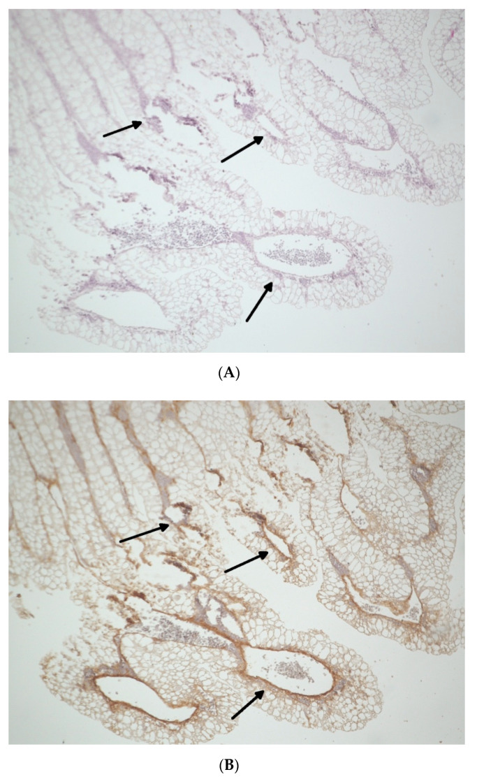
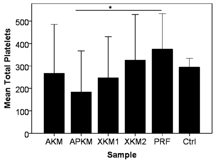
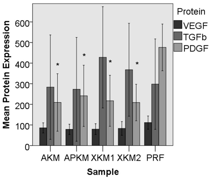
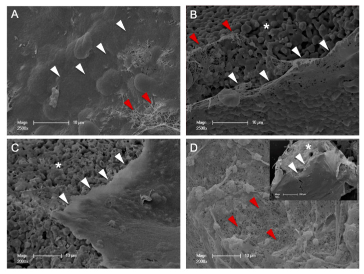
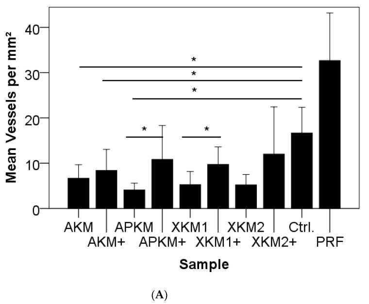
The impaired angiogenic potential of bone substitute materials (BSMs) may limit regenerative processes. Therefore, changes in the angiogenetic properties of different BSMs in combination with platelet-rich fibrin (PRF) in comparison to PRF alone, as well as to native BSMs, were analyzed in vitro and in vivo to evaluate possible clinical application. In vitro, four BSMs of different origins (allogeneic, alloplastic, and xenogeneic) were biofunctionalized with PRF and compared to PRF in terms of platelet interaction and growth factor release (vascular endothelial growth factor (VEGF), tissue growth factor ß (TGFß) and platelet-derived growth factor (PDGF)) after 15 min. To visualize initial cell-cell interactions, SEM was performed. In vivo, all BSMs (±PRF) were analyzed after 24 h for new-formed vessels using a chorioallantoic membrane (CAM) assay. Especially for alloplastic BSMs, the addition of PRF led to a significant consumption of platelets ( = 0.05). PDGF expression significantly decreased in comparison to PRF alone (all BSMs: < 0.013). SEM showed the close spatial relation of each BSM and PRF. In vivo, PRF had a significant positive pro-angiogenic influence in combination with alloplastic ( = 0.007) and xenogeneic materials ( = 0.015) in comparison to the native BSMs. For bio-activated xenogeneic BSMs, the branching points were also significantly increased ( = 0.005). Finally, vessel formation was increased for BSMs and PRF in comparison to the native control (allogeneic: = 0.046; alloplastic: = 0.046; and xenogeneic: = 0.050). An early enhancement of angiogenetic properties was demonstrated when combining BSMs with PRF in vitro and led to upregulated vessel formation in vivo. Thus, the use of BSMs in combination with PRF may trigger bony regeneration in clinical approaches.
骨替代材料(BSMs)血管生成潜力受损可能会限制再生过程。因此,为评估不同BSMs与富血小板纤维蛋白(PRF)联合使用相对于单独使用PRF以及天然BSMs时血管生成特性的变化,进行了体外和体内分析以评估其可能的临床应用。在体外,四种不同来源(同种异体、人工合成和异种)的BSMs用PRF进行生物功能化处理,并在15分钟后就血小板相互作用和生长因子释放(血管内皮生长因子(VEGF)、组织生长因子β(TGFβ)和血小板衍生生长因子(PDGF))方面与PRF进行比较。为观察初始细胞间相互作用,进行了扫描电子显微镜(SEM)检查。在体内,使用尿囊绒膜(CAM)试验在24小时后分析所有BSMs(±PRF)的新生血管情况。特别是对于人工合成BSMs,添加PRF导致血小板显著消耗(P = 0.05)。与单独使用PRF相比,PDGF表达显著降低(所有BSMs:P < 0.013)。SEM显示每种BSM与PRF之间存在紧密的空间关系。在体内,与天然BSMs相比,PRF与人工合成材料(P = 0.007)和异种材料联合使用时具有显著的促血管生成的积极影响(P = 0.015)。对于生物活化的异种BSMs,分支点也显著增加(P = 0.005)。最后,与天然对照相比,BSMs和PRF联合使用时血管形成增加(同种异体:P = 0.046;人工合成:P = 0.046;异种:P = 0.050)。体外将BSMs与PRF联合使用时证明血管生成特性早期增强,并导致体内血管形成上调。因此,在临床方法中使用BSMs与PRF联合可能会触发骨再生。