The Wistar Institute, Philadelphia, PA, USA.
Division of Cancer Epidemiology and Genetics, National Cancer Institute, Bethesda, MD, USA.
Nat Commun. 2021 Jan 12;12(1):346. doi: 10.1038/s41467-020-20600-7.
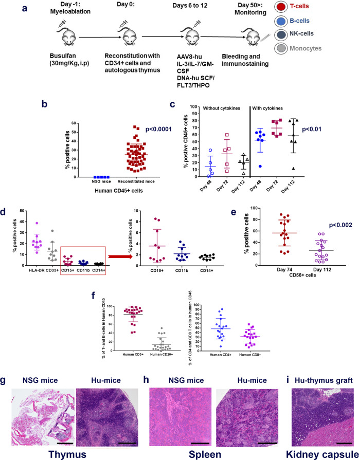
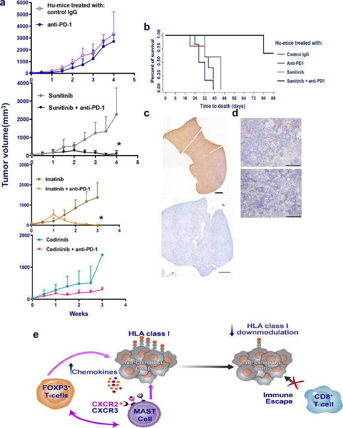
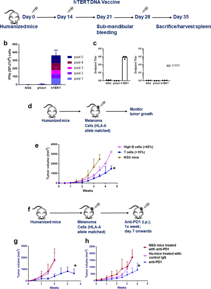
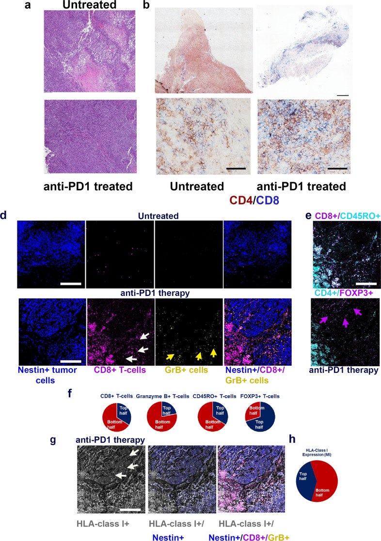
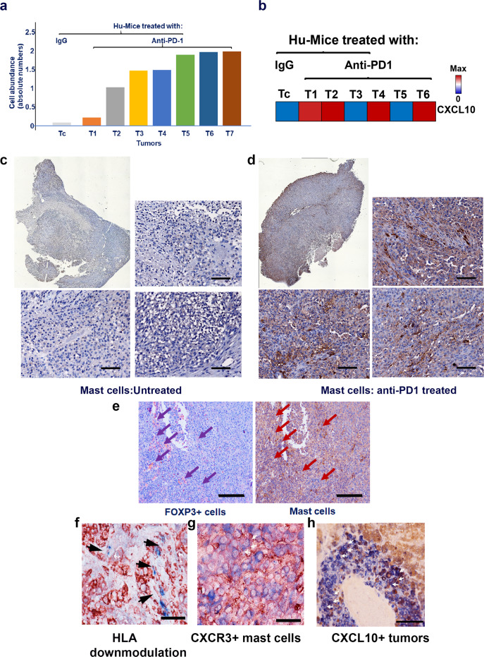
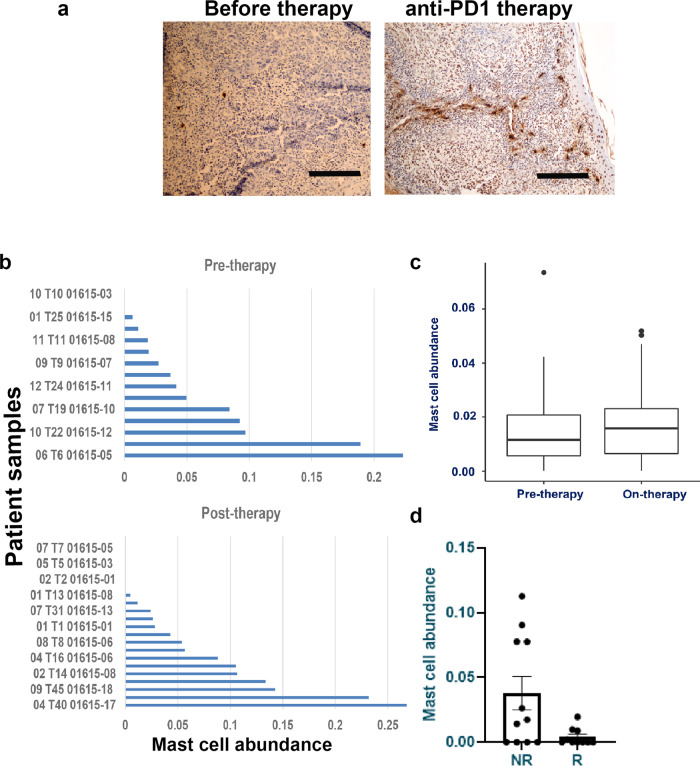
Anti-PD-1 therapy is used as a front-line treatment for many cancers, but mechanistic insight into this therapy resistance is still lacking. Here we generate a humanized (Hu)-mouse melanoma model by injecting fetal liver-derived CD34 cells and implanting autologous thymus in immune-deficient NOD-scid IL2Rγ (NSG) mice. Reconstituted Hu-mice are challenged with HLA-matched melanomas and treated with anti-PD-1, which results in restricted tumor growth but not complete regression. Tumor RNA-seq, multiplexed imaging and immunohistology staining show high expression of chemokines, as well as recruitment of FOXP3 Treg and mast cells, in selective tumor regions. Reduced HLA-class I expression and CD8/Granz B T cells homeostasis are observed in tumor regions where FOXP3 Treg and mast cells co-localize, with such features associated with resistance to anti-PD-1 treatment. Combining anti-PD-1 with sunitinib or imatinib results in the depletion of mast cells and complete regression of tumors. Our results thus implicate mast cell depletion for improving the efficacy of anti-PD-1 therapy.
抗 PD-1 疗法被用作许多癌症的一线治疗方法,但对这种治疗耐药性的机制理解仍很缺乏。在这里,我们通过注射胎肝来源的 CD34 细胞并在免疫缺陷 NOD-scid IL2Rγ(NSG)小鼠中植入自体胸腺来生成人源化(Hu)-小鼠黑色素瘤模型。重建的 Hu-小鼠接受与 HLA 匹配的黑色素瘤的挑战,并接受抗 PD-1 治疗,这导致肿瘤生长受限但不完全消退。肿瘤 RNA-seq、多重成像和免疫组织化学染色显示趋化因子高表达,以及 FOXP3 Treg 和肥大细胞在选择性肿瘤区域的募集。在 FOXP3 Treg 和肥大细胞共定位的肿瘤区域观察到 HLA 类 I 表达和 CD8/Granz B T 细胞稳态减少,这些特征与抗 PD-1 治疗的耐药性相关。联合使用抗 PD-1 和舒尼替尼或伊马替尼可导致肥大细胞耗竭并使肿瘤完全消退。因此,我们的结果表明,肥大细胞耗竭可提高抗 PD-1 治疗的疗效。