Cardiology Clinic, "Alexandrovska" University Hospital, Medical University of Sofia, Bulgaria.
Cardiol J. 2023;30(2):221-227. doi: 10.5603/CJ.a2021.0003. Epub 2021 Jan 13.
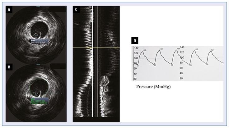
Aortic stiffness is a well-known cardio-vascular risk factor. For years, different methods have been studied in the assessment of aortic elastic properties and large arterial stiffness for risk stratification. Herein is an assessment of the role of intravascular ultrasound (IVUS) imaging for the evaluation of aortic elastic properties.
Intravascular ultrasound imaging of the aorta was performed in 12 patients with transthoracic echocardiography (TTE) and computed tomography (CT) evidence for enlargement of the ascending aorta - diameter ≥ 40.0 mm. Mechanical properties of the aorta were derived from the measured diameters and intra-aortic pressure. Paired samples T-test analyses were performed to determine differences between measurements derived by TTE, CT and IVUS.
Mean values of the calculated elastic properties via IVUS of the ascending aorta were as follows: compliance 0.021 ± 0.02; strain 205 ± 4.3; aortic stiffness index 4.3 ± 0.75; elastic modulus 0.31 ± 0.05. On paired T-test analysis maximum ascending aortic diameter measured by CT aortography and IVUS did not differ significantly (t = -0.19, p = 0.985), but a significant difference between IVUS measurements and TTE derived diameters was found (t = 13.118, p = 0.034). On average, IVUS diameters were 2.3 mm larger than the results acquired by TTE (95% confidence interval: 14.21-17.13).
Intravascular ultrasound examination of the ascending aorta provided larger diameters than the ones collected by means of TTE. However, IVUS measurements did not differ significantly from diameters derived by CT aortography.
主动脉僵硬是众所周知的心血管风险因素。多年来,人们一直在研究不同的方法来评估主动脉弹性特性和大动脉僵硬程度,以进行风险分层。本文评估了血管内超声(IVUS)成像在评估主动脉弹性特性中的作用。
对 12 例经胸超声心动图(TTE)和计算机断层扫描(CT)证实升主动脉扩张的患者进行了主动脉 IVUS 成像 - 直径≥40.0mm。通过测量的直径和主动脉内压来推导出主动脉的力学特性。采用配对样本 T 检验分析来确定 TTE、CT 和 IVUS 测量值之间的差异。
通过 IVUS 计算得出的升主动脉弹性特性的平均值如下:顺应性 0.021±0.02;应变 205±4.3;主动脉僵硬指数 4.3±0.75;弹性模量 0.31±0.05。在配对 T 检验分析中,CT 血管造影和 IVUS 测量的升主动脉最大直径无显著差异(t=-0.19,p=0.985),但 IVUS 测量值与 TTE 衍生直径之间存在显著差异(t=13.118,p=0.034)。平均而言,IVUS 直径比 TTE 获得的结果大 2.3mm(95%置信区间:14.21-17.13)。
升主动脉的血管内超声检查提供的直径大于 TTE 采集的直径。然而,IVUS 测量值与 CT 血管造影得出的直径无显著差异。