Department of Medical Imaging, Guangdong Traditional Medical and Sports Injury Rehabilitation Research Institute, Guangdong Second Provincial General Hospital, Haizhu District, Shiliugang Rd, Guangzhou, 510317, People's Republic of China.
Department of Medical Imaging, Fancheng District, Xiangyang Central Hospital, Affiliated Hospital of Hubei University of Arts and Science, Zhongyuan RD, Xiangyang, 441003, People's Republic of China.
Sci Rep. 2021 Jan 13;11(1):1103. doi: 10.1038/s41598-020-79183-4.
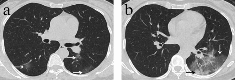
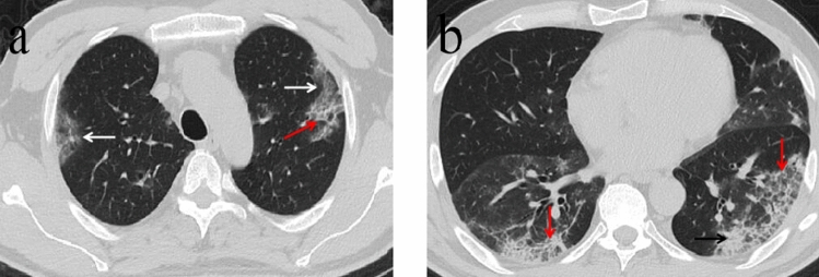
The aim of this study was to analyze initial chest computed tomography (CT) findings in COVID-19 pneumonia and identify features associated with poor prognosis. Patients with RT-PCR-confirmed COVID-19 infection were assigned to recovery group if they made a full recovery and to death group if they died within 2 months of hospitalization. Chest CT examinations for ground-glass opacity, crazy-paving pattern, consolidation, and fibrosis were scored by two reviewers. The total CT score comprised the sum of lung involvement (5 lobes, scores 1-5 for each lobe, range; 0, none; 25, maximum). 40 patients who recovered from COVID-19 and six patients who died were enrolled. The initial chest CTs showed 27 (58.7%) patients had ground-glass opacity, 19 (41.3%) had ground glass and consolidation, and 35 (76.1%) patients had crazy-paving pattern. None of the patients who died had fibrosis in contrast to six (15%) patients who recovered from COVID-19. Most patients had subpleural lesions (89.0%) as well as bilateral (87.0%) and lower (93.0%) lung lobe involvement. Diffuse lesions were present in four (67%) patients who succumbed to coronavirus but only one (2.5%) patient who recovered (p < 0.001). In the death group of patients, the total CT score was higher than that of the recovery group (p = 0.005). Patients in the death group had lower lymphocyte count and higher C-reactive protein than those in the recovery group (p = 0.011 and p = 0.041, respectively). A high CT score and diffuse distribution of lung lesions in COVID-19 are indicative of disease severity and short-term mortality.
本研究旨在分析 COVID-19 肺炎的初始胸部计算机断层扫描(CT)结果,并确定与不良预后相关的特征。经 RT-PCR 确诊为 COVID-19 感染的患者,如果在住院后 2 个月内完全康复,则归入康复组,如果死亡,则归入死亡组。两名审阅者对磨玻璃影、铺路石征、实变和纤维化的胸部 CT 检查进行评分。总 CT 评分由肺受累程度(5 个肺叶,每个肺叶评分为 1-5 分,范围为 0 分,无;25 分,最高分)组成。共纳入 40 例从 COVID-19 中康复的患者和 6 例死亡的患者。最初的胸部 CT 显示 27 例(58.7%)患者有磨玻璃影,19 例(41.3%)有磨玻璃影和实变,35 例(76.1%)患者有铺路石征。与从 COVID-19 中康复的 6 例(15%)患者相比,死亡患者无一例有纤维化。大多数患者均有胸膜下病变(89.0%)、双侧(87.0%)和下肺叶受累(93.0%)。4 例(67%)死于冠状病毒的患者有弥漫性病变,但只有 1 例(2.5%)康复患者有弥漫性病变(p<0.001)。在死亡组患者中,总 CT 评分高于康复组(p=0.005)。死亡组患者的淋巴细胞计数和 C 反应蛋白均低于康复组(p=0.011 和 p=0.041)。COVID-19 中高 CT 评分和弥漫性肺病变提示疾病严重程度和短期死亡率高。