Relucenti Michela, Familiari Giuseppe, Donfrancesco Orlando, Taurino Maurizio, Li Xiaobo, Chen Rui, Artini Marco, Papa Rosanna, Selan Laura
Department of Anatomy, Histology, Forensic Medicine and Orthopedics, Sapienza University of Rome, Via Alfonso Borelli 50, 00161 Rome, Italy.
Department of Clinical and Molecular Medicine, Unit of Vascular Surgery, Sant'Andrea Hospital, Sapienza University of Rome, Via di Grottarossa 1039, 00189 Rome, Italy.
Biology (Basel). 2021 Jan 12;10(1):51. doi: 10.3390/biology10010051.
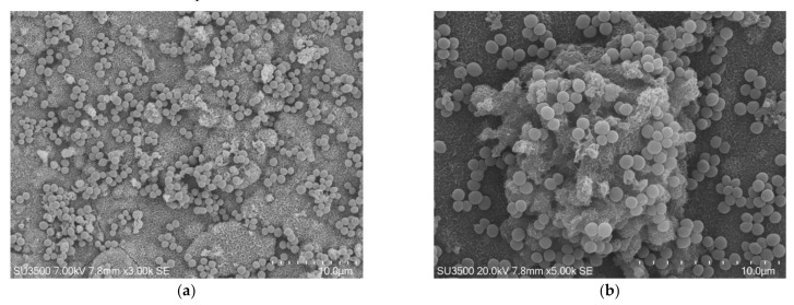
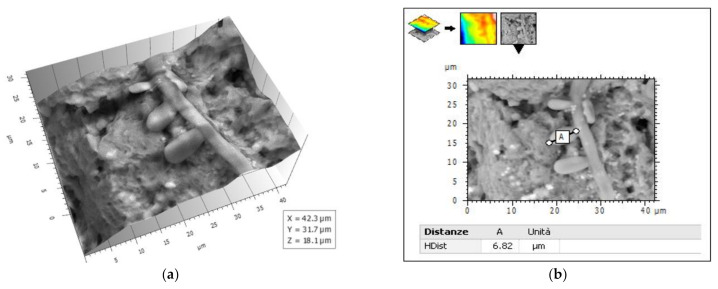
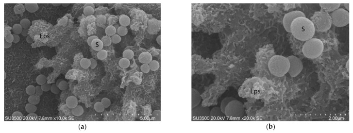
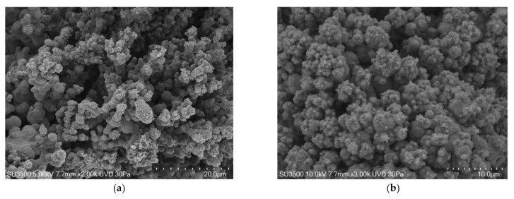
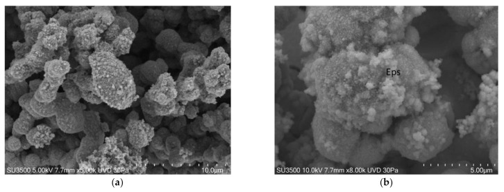
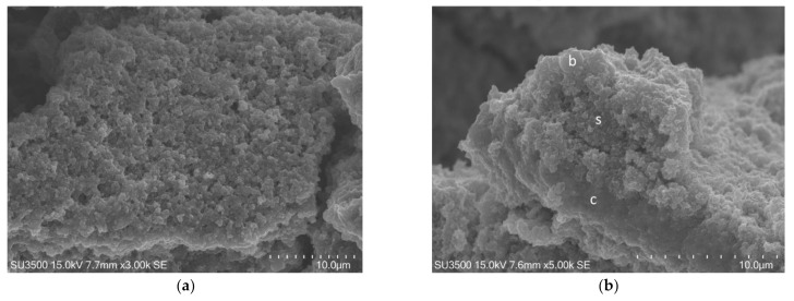
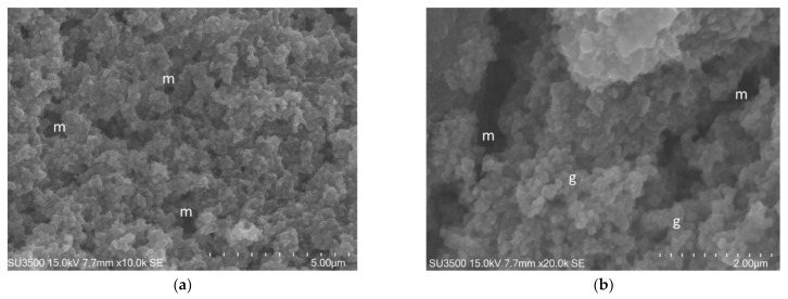
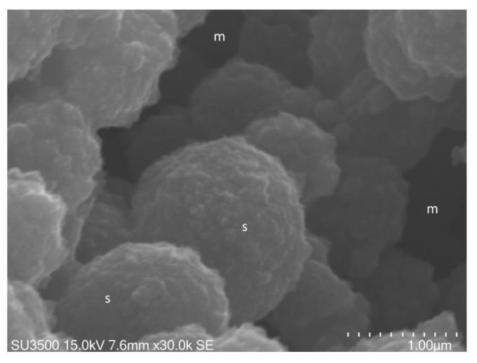
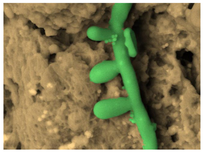
Several imaging methodologies have been used in biofilm studies, contributing to deepening the knowledge on their structure. This review illustrates the most widely used microscopy techniques in biofilm investigations, focusing on traditional and innovative scanning electron microscopy techniques such as scanning electron microscopy (SEM), variable pressure SEM (VP-SEM), environmental SEM (ESEM), and the more recent ambiental SEM (ASEM), ending with the cutting edge Cryo-SEM and focused ion beam SEM (FIB SEM), highlighting the pros and cons of several methods with particular emphasis on conventional SEM and VP-SEM. As each technique has its own advantages and disadvantages, the choice of the most appropriate method must be done carefully, based on the specific aim of the study. The evaluation of the drug effects on biofilm requires imaging methods that show the most detailed ultrastructural features of the biofilm. In this kind of research, the use of scanning electron microscopy with customized protocols such as osmium tetroxide (OsO), ruthenium red (RR), tannic acid (TA) staining, and ionic liquid (IL) treatment is unrivalled for its image quality, magnification, resolution, minimal sample loss, and actual sample structure preservation. The combined use of innovative SEM protocols and 3-D image analysis software will allow for quantitative data from SEM images to be extracted; in this way, data from images of samples that have undergone different antibiofilm treatments can be compared.
几种成像方法已用于生物膜研究,有助于加深对其结构的了解。本综述阐述了生物膜研究中最常用的显微镜技术,重点介绍传统和创新的扫描电子显微镜技术,如扫描电子显微镜(SEM)、可变压力扫描电子显微镜(VP-SEM)、环境扫描电子显微镜(ESEM)以及最新的常压扫描电子显微镜(ASEM),最后介绍前沿的低温扫描电子显微镜和聚焦离子束扫描电子显微镜(FIB SEM),强调了几种方法的优缺点,尤其着重介绍了传统扫描电子显微镜和可变压力扫描电子显微镜。由于每种技术都有其优缺点,必须根据研究的具体目的谨慎选择最合适的方法。评估药物对生物膜的作用需要能展示生物膜最详细超微结构特征的成像方法。在这类研究中,使用诸如四氧化锇(OsO)、钌红(RR)、单宁酸(TA)染色和离子液体(IL)处理等定制方案的扫描电子显微镜,在图像质量、放大倍数、分辨率、最小样本损失和实际样本结构保存方面无与伦比。创新的扫描电子显微镜方案与三维图像分析软件的结合使用将能够提取扫描电子显微镜图像中的定量数据;通过这种方式,可以比较经过不同抗生物膜处理的样本图像数据。