Inserm U1259 MAVIVH, Université de Tours and CHRU de Tours, Tours, France.
Plate-Forme IBiSA de Microscopie Electronique, Université de Tours and CHRU de Tours, Tours, France.
Cell Mol Life Sci. 2021 Apr;78(7):3565-3576. doi: 10.1007/s00018-020-03745-y. Epub 2021 Jan 15.
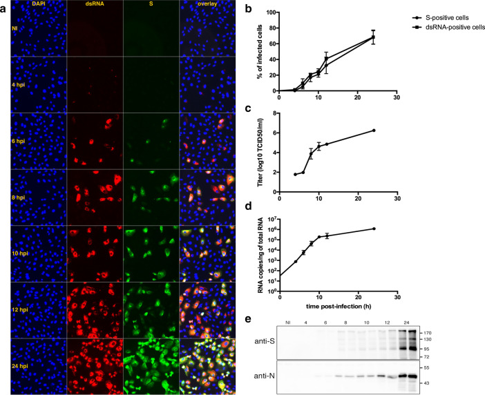
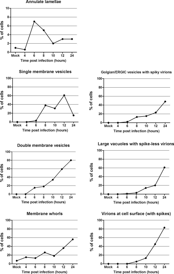
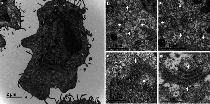
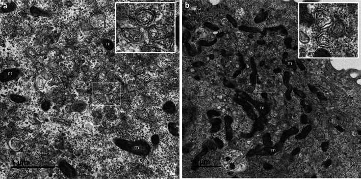
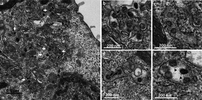
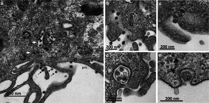
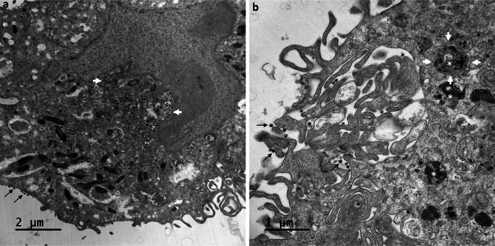
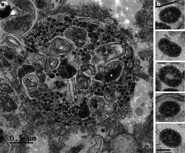
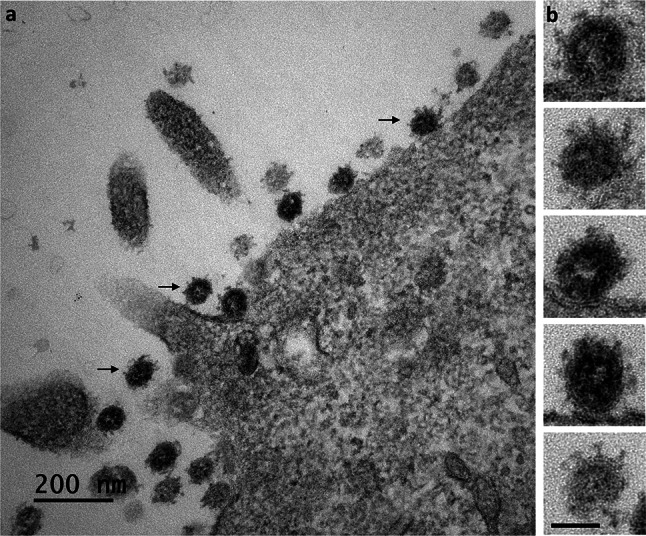
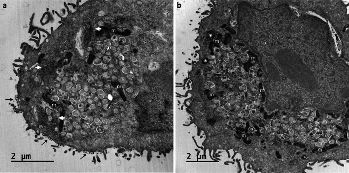
Many studies on SARS-CoV-2 have been performed over short-time scale, but few have focused on the ultrastructural characteristics of infected cells. We used TEM to perform kinetic analysis of the ultrastructure of SARS-CoV-2-infected cells. Early infection events were characterized by the presence of clusters of single-membrane vesicles and stacks of membrane containing nuclear pores called annulate lamellae (AL). A large network of host cell-derived organelles transformed into virus factories was subsequently observed in the cells. As previously described for other RNA viruses, these replication factories consisted of double-membrane vesicles (DMVs) located close to the nucleus. Viruses released at the cell surface by exocytosis harbored the typical crown of spike proteins, but viral particles without spikes were also observed in intracellular compartments, possibly reflecting incorrect assembly or a cell degradation process.
许多关于 SARS-CoV-2 的研究都是在短时间尺度上进行的,但很少有研究关注感染细胞的超微结构特征。我们使用 TEM 对 SARS-CoV-2 感染细胞的超微结构进行了动力学分析。早期感染事件的特征是存在单个膜泡簇和称为环状层板(annulate lamellae,AL)的含有核孔的膜堆栈。随后在细胞中观察到大量由宿主细胞衍生的细胞器转化为病毒工厂。与其他 RNA 病毒一样,这些复制工厂由靠近细胞核的双层膜囊泡(double-membrane vesicles,DMVs)组成。通过胞吐作用释放到细胞表面的病毒颗粒带有典型的刺突蛋白冠状,但也在细胞内隔室中观察到没有刺突的病毒颗粒,这可能反映了不正确的组装或细胞降解过程。