Bordoloi Bharadwaj, Siddiqui Safia, Jaiswal Rohit, Tandon Aanchal, Jain Amol, Chaturvedi Rashmi
Department of Dentistry, Garmur SDCH, Majuli, Assam, India.
Department of Oral Pathology and Microbiology, Sardar Patel Postgraduate Institute of Dental and Medical Sciences, Lucknow, Uttar Pradesh, India.
J Oral Maxillofac Pathol. 2020 May-Aug;24(2):398. doi: 10.4103/jomfp.JOMFP_84_18. Epub 2020 Sep 9.
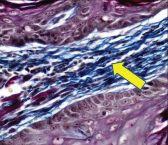
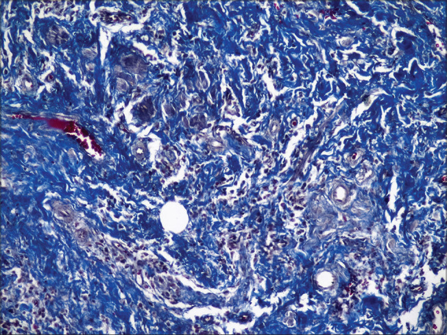
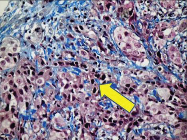
Solid tumors such as oral squamous cell carcinoma (OSCC) are composed of malignant epithelial cells and the stroma in which these cells are dispersed. As the tumor progresses, the extracellular matrix undergoes dramatic morphological and architectural changes. Special stains make analysis easy and less erroneous by highlighting the area of interest and can be used to study these changes.
The aim of the study was to analyze morphological changes in collagen fibers in various histological grades of OSCC using Masson's trichrome (MT) and Picrosirius red (PSR).
The study comprised 74 tissue samples, divided into two groups: Group I consisted of 63 cases of histologically proven OSCC (39 cases of well-differentiated squamous cell carcinoma [WDSCC], 17 moderately differentiated squamous cell carcinoma [MDSCC] and 7 poorly differentiated squamous cell carcinoma [PDSCC]) and Group II consisted of 11 cases of normal mucosa as controls.
Sections were stained with hematoxylin and eosin, MT and PSR and observed under light and polarizing microscope, respectively.
ANOVA, Tukey's honestly significant difference multiple comparison test, Chi-square test and paired -test were used for the statistical analysis.
As the grade of OSCC progressed, collagen fibers became thin, loosely packed and haphazard. The mean area fraction also decreased. They exhibited orange-red hue and strong birefringence in WDSCC, yellowish-orange hue and strong birefringence in MDSCC and greenish-yellow hue and weak birefringence in PDSCC.
Initially, there is a reorganization of the collagen fibers in an attempt to prevent the invasion of tumor cells, but as cancer progresses, the stromal change enhances movement of the tumor cells within it, leading to metastasis.
实体瘤,如口腔鳞状细胞癌(OSCC),由恶性上皮细胞和这些细胞散布其中的基质组成。随着肿瘤进展,细胞外基质会经历显著的形态和结构变化。特殊染色通过突出感兴趣的区域使分析变得容易且减少错误,可用于研究这些变化。
本研究旨在使用马松三色染色法(MT)和天狼星红苦味酸染色法(PSR)分析不同组织学分级的OSCC中胶原纤维的形态变化。
本研究包括74个组织样本,分为两组:第一组由63例经组织学证实的OSCC病例组成(39例高分化鳞状细胞癌[WDSCC]、17例中分化鳞状细胞癌[MDSCC]和7例低分化鳞状细胞癌[PDSCC]),第二组由11例正常黏膜作为对照组成。
切片分别用苏木精和伊红、MT和PSR染色,并分别在光学显微镜和偏光显微镜下观察。
采用方差分析、Tukey真实显著差异多重比较检验、卡方检验和配对检验进行统计分析。
随着OSCC分级的进展,胶原纤维变细、排列疏松且杂乱无章。平均面积分数也降低。它们在WDSCC中呈现橙红色调且双折射强,在MDSCC中呈现黄橙色调且双折射强,在PDSCC中呈现绿黄色调且双折射弱。
最初,胶原纤维会进行重新组织以试图阻止肿瘤细胞的侵袭,但随着癌症进展,基质变化会增强肿瘤细胞在其中的移动,导致转移。