• 文献检索
  • 文档翻译
  • 深度研究
  • 学术资讯
  • Suppr Zotero 插件Zotero 插件
  • 邀请有礼
  • 套餐&价格
  • 历史记录
应用&插件
Suppr Zotero 插件Zotero 插件浏览器插件Mac 客户端Windows 客户端微信小程序
定价
高级版会员购买积分包购买API积分包
服务
文献检索文档翻译深度研究API 文档MCP 服务
关于我们
关于 Suppr公司介绍联系我们用户协议隐私条款
关注我们

Suppr 超能文献

核心技术专利:CN118964589B侵权必究
粤ICP备2023148730 号-1Suppr @ 2026

文献检索

告别复杂PubMed语法,用中文像聊天一样搜索,搜遍4000万医学文献。AI智能推荐,让科研检索更轻松。

立即免费搜索

文件翻译

保留排版,准确专业,支持PDF/Word/PPT等文件格式,支持 12+语言互译。

免费翻译文档

深度研究

AI帮你快速写综述,25分钟生成高质量综述,智能提取关键信息,辅助科研写作。

立即免费体验

使用磁共振成像测量计划与质子束治疗位置之间的眼球变形。

Measuring eye deformation between planning and proton beam therapy position using magnetic resonance imaging.

作者信息

Jaarsma-Coes Myriam G, Marinkovic Marina, Astreinidou Eleftheria, Schuurmans Megan S, Peters Femke P, Luyten Gregorius P M, Rasch Coen R N, Beenakker Jan-Willem M

机构信息

Ophthalmology, Leiden University Medical Centre, Leiden, Netherlands.

Radiology, C.J. Gorter Centre for High Field MRI, Leiden University Medical Centre, Leiden, Netherlands.

出版信息

Phys Imaging Radiat Oncol. 2020 Oct 6;16:33-36. doi: 10.1016/j.phro.2020.09.010. eCollection 2020 Oct.

DOI:10.1016/j.phro.2020.09.010
PMID:33458341
原文链接:https://pmc.ncbi.nlm.nih.gov/articles/PMC7807689/
Abstract

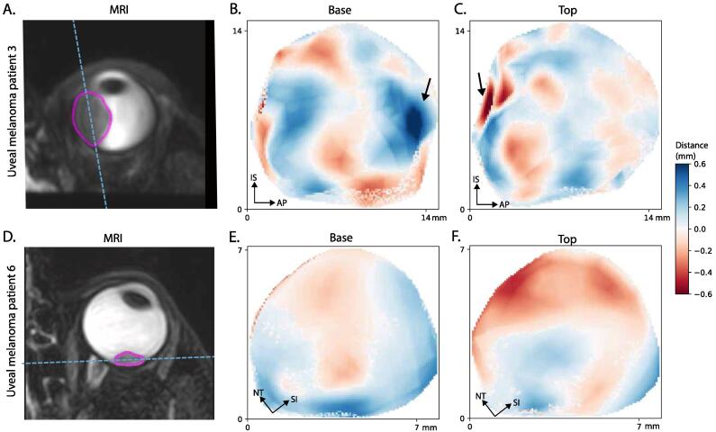
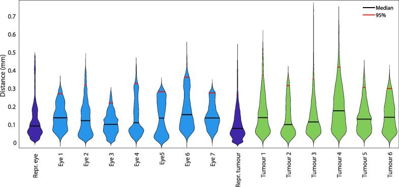
Proton beam therapy (PBT) for uveal melanoma (UM) is performed in sitting position, while the acquisition of the Magnetic resonance (MR)-images for treatment planning is performed in supine position. We assessed the effect of this difference in position on the eye- and tumour- shape. Seven subjects and six UM-patients were scanned in supine and a seating mimicking position. The distances between the tumour/sclera in both positions were calculated. The median distance between both positions was 0.1 mm. Change in gravity direction produced no substantial changes in sclera and tumour shape, indicating that supinely acquired MR-images can be used to plan ocular-PBT.

摘要

葡萄膜黑色素瘤(UM)的质子束治疗(PBT)是在坐姿下进行的,而用于治疗计划的磁共振(MR)图像采集则是在仰卧位进行的。我们评估了这种体位差异对眼睛和肿瘤形状的影响。对7名受试者和6名UM患者在仰卧位和模拟坐姿下进行了扫描。计算了两个体位下肿瘤/巩膜之间的距离。两个体位之间的中位距离为0.1毫米。重力方向的改变未导致巩膜和肿瘤形状发生实质性变化,这表明仰卧位采集的MR图像可用于眼部PBT的治疗计划。

https://cdn.ncbi.nlm.nih.gov/pmc/blobs/d81e/7807689/ac02b7daa8c4/gr2.jpg
https://cdn.ncbi.nlm.nih.gov/pmc/blobs/d81e/7807689/d72ea9d912ed/gr1.jpg
https://cdn.ncbi.nlm.nih.gov/pmc/blobs/d81e/7807689/ac02b7daa8c4/gr2.jpg
https://cdn.ncbi.nlm.nih.gov/pmc/blobs/d81e/7807689/d72ea9d912ed/gr1.jpg
https://cdn.ncbi.nlm.nih.gov/pmc/blobs/d81e/7807689/ac02b7daa8c4/gr2.jpg

相似文献

1
Measuring eye deformation between planning and proton beam therapy position using magnetic resonance imaging.使用磁共振成像测量计划与质子束治疗位置之间的眼球变形。
Phys Imaging Radiat Oncol. 2020 Oct 6;16:33-36. doi: 10.1016/j.phro.2020.09.010. eCollection 2020 Oct.
2
The Economic Value of MR-Imaging for Uveal Melanoma.磁共振成像对葡萄膜黑色素瘤的经济价值
Clin Ophthalmol. 2020 Apr 28;14:1135-1143. doi: 10.2147/OPTH.S238405. eCollection 2020.
3
Comparison of Magnetic Resonance Imaging-Based and Conventional Measurements for Proton Beam Therapy of Uveal Melanoma.基于磁共振成像的与常规测量在葡萄膜黑色素瘤质子束治疗中的比较。
Ophthalmol Retina. 2023 Feb;7(2):178-188. doi: 10.1016/j.oret.2022.06.019. Epub 2022 Jul 13.
4
Personalized Anatomic Eye Model From T1-Weighted Volume Interpolated Gradient Echo Magnetic Resonance Imaging of Patients With Uveal Melanoma.基于 T1 加权容积内插梯度回波磁共振成像的葡萄膜黑色素瘤患者个性化解剖眼球模型。
Int J Radiat Oncol Biol Phys. 2018 Nov 15;102(4):813-820. doi: 10.1016/j.ijrobp.2018.05.004. Epub 2018 May 14.
5
Uveal melanoma: quantitative evaluation of diffusion-weighted MR imaging in the response assessment after proton-beam therapy, long-term follow-up.葡萄膜黑色素瘤:质子束治疗后反应评估中扩散加权磁共振成像的定量评估,长期随访
Radiol Med. 2017 Feb;122(2):131-139. doi: 10.1007/s11547-016-0697-3. Epub 2016 Oct 17.
6
Neoadjuvant proton beam irradiation vs. adjuvant ruthenium brachytherapy in transscleral resection of uveal melanoma.新辅助质子束照射与辅助钌近距离放射治疗在葡萄膜黑色素瘤经巩膜切除术中的比较
Graefes Arch Clin Exp Ophthalmol. 2018 Sep;256(9):1767-1775. doi: 10.1007/s00417-018-4032-7. Epub 2018 Jun 16.
7
Clinical evaluation of ultra-high-field MRI for three-dimensional visualisation of tumour size in uveal melanoma patients, with direct relevance to treatment planning.超高场磁共振成像用于葡萄膜黑色素瘤患者肿瘤大小三维可视化的临床评估,与治疗规划直接相关。
MAGMA. 2016 Jun;29(3):571-7. doi: 10.1007/s10334-016-0529-4. Epub 2016 Feb 25.
8
Use of high-frequency ultrasound imaging to improve delineation of anterior uveal melanoma for proton irradiation.使用高频超声成像改善质子放疗前葡萄膜黑色素瘤的轮廓描绘。
Phys Med Biol. 2001 Feb;46(2):579-90. doi: 10.1088/0031-9155/46/2/322.
9
Practice Patterns Analysis of Ocular Proton Therapy Centers: The International OPTIC Survey.眼质子治疗中心实践模式分析:国际 OPTIC 调查。
Int J Radiat Oncol Biol Phys. 2016 May 1;95(1):336-343. doi: 10.1016/j.ijrobp.2016.01.040. Epub 2016 Jan 28.
10
3D MRI-based tumor delineation of ocular melanoma and its comparison with conventional techniques.基于3D磁共振成像的眼内黑色素瘤肿瘤轮廓描绘及其与传统技术的比较。
Med Phys. 2005 Nov;32(11):3355-62. doi: 10.1118/1.2068927.

引用本文的文献

1
Magnetic Resonance Imaging in the Clinical Care for Uveal Melanoma Patients-A Systematic Review from an Ophthalmic Perspective.从眼科视角对葡萄膜黑色素瘤患者临床护理中磁共振成像的系统评价
Cancers (Basel). 2023 May 30;15(11):2995. doi: 10.3390/cancers15112995.
2
Inter-Observer Variability in MR-Based Target Volume Delineation of Uveal Melanoma.基于磁共振成像的葡萄膜黑色素瘤靶区勾画的观察者间变异性
Adv Radiat Oncol. 2022 Dec 24;8(3):101149. doi: 10.1016/j.adro.2022.101149. eCollection 2023 May-Jun.
3
Clinical and imaging clues to the diagnosis and follow-up of ptosis and ophthalmoparesis.

本文引用的文献

1
MRI of Uveal Melanoma.葡萄膜黑色素瘤的磁共振成像
Cancers (Basel). 2019 Mar 17;11(3):377. doi: 10.3390/cancers11030377.
2
Can CT imaging improve targeting accuracy in clip-based proton therapy of ocular melanoma?CT 成像能否提高基于夹式质子治疗眼黑色素瘤的靶向准确性?
Phys Med Biol. 2019 Jan 24;64(3):035010. doi: 10.1088/1361-6560/aaf9c9.
3
Personalized Anatomic Eye Model From T1-Weighted Volume Interpolated Gradient Echo Magnetic Resonance Imaging of Patients With Uveal Melanoma.基于 T1 加权容积内插梯度回波磁共振成像的葡萄膜黑色素瘤患者个性化解剖眼球模型。
眼睑下垂和眼肌瘫痪的诊断和随访的临床及影像学线索。
J Cachexia Sarcopenia Muscle. 2022 Dec;13(6):2820-2834. doi: 10.1002/jcsm.13089. Epub 2022 Sep 29.
4
Histopathologic and MR Imaging Appearance of Spontaneous and Radiation-Induced Necrosis in Uveal Melanomas: Initial Results.葡萄膜黑色素瘤中自发性坏死与放射性坏死的组织病理学及磁共振成像表现:初步结果
Cancers (Basel). 2022 Jan 2;14(1):215. doi: 10.3390/cancers14010215.
5
Characterization of the HollandPTC proton therapy beamline dedicated to uveal melanoma treatment and an interinstitutional comparison.用于脉络膜黑色素瘤治疗的 HollandPTC 质子治疗束线的特性描述及机构间比较。
Med Phys. 2021 Aug;48(8):4506-4522. doi: 10.1002/mp.15024. Epub 2021 Jul 11.
Int J Radiat Oncol Biol Phys. 2018 Nov 15;102(4):813-820. doi: 10.1016/j.ijrobp.2018.05.004. Epub 2018 May 14.
4
Uveal melanoma: epidemiology, etiology, and treatment of primary disease.葡萄膜黑色素瘤:原发性疾病的流行病学、病因学及治疗
Clin Ophthalmol. 2017 Jan 31;11:279-289. doi: 10.2147/OPTH.S89591. eCollection 2017.
5
Practice Patterns Analysis of Ocular Proton Therapy Centers: The International OPTIC Survey.眼质子治疗中心实践模式分析:国际 OPTIC 调查。
Int J Radiat Oncol Biol Phys. 2016 May 1;95(1):336-343. doi: 10.1016/j.ijrobp.2016.01.040. Epub 2016 Jan 28.
6
Clinical Outcomes of Proton Radiotherapy for Uveal Melanoma.葡萄膜黑色素瘤质子放疗的临床结果
Clin Oncol (R Coll Radiol). 2016 Aug;28(8):e17-27. doi: 10.1016/j.clon.2016.01.034. Epub 2016 Feb 23.
7
Clinical evaluation of ultra-high-field MRI for three-dimensional visualisation of tumour size in uveal melanoma patients, with direct relevance to treatment planning.超高场磁共振成像用于葡萄膜黑色素瘤患者肿瘤大小三维可视化的临床评估,与治疗规划直接相关。
MAGMA. 2016 Jun;29(3):571-7. doi: 10.1007/s10334-016-0529-4. Epub 2016 Feb 25.
8
Clinical Management of Uveal and Conjunctival Melanoma.葡萄膜和结膜黑色素瘤的临床管理
Oncology (Williston Park). 2016 Jan;30(1):29-32, 34-43, 48.
9
Outcomes of treatment with stereotactic radiosurgery or proton beam therapy for choroidal melanoma.立体定向放射外科或质子束治疗脉络膜黑色素瘤的治疗结果。
Eye (Lond). 2015 Sep;29(9):1194-8. doi: 10.1038/eye.2015.109. Epub 2015 Jul 10.
10
Eye tracking and gating system for proton therapy of orbital tumors.眼球跟踪和门控系统在眼眶肿瘤质子治疗中的应用。
Med Phys. 2012 Jul;39(7):4265-73. doi: 10.1118/1.4729708.