• 文献检索
  • 文档翻译
  • 深度研究
  • 学术资讯
  • Suppr Zotero 插件Zotero 插件
  • 邀请有礼
  • 套餐&价格
  • 历史记录
应用&插件
Suppr Zotero 插件Zotero 插件浏览器插件Mac 客户端Windows 客户端微信小程序
定价
高级版会员购买积分包购买API积分包
服务
文献检索文档翻译深度研究API 文档MCP 服务
关于我们
关于 Suppr公司介绍联系我们用户协议隐私条款
关注我们

Suppr 超能文献

核心技术专利:CN118964589B侵权必究
粤ICP备2023148730 号-1Suppr @ 2026

文献检索

告别复杂PubMed语法,用中文像聊天一样搜索,搜遍4000万医学文献。AI智能推荐,让科研检索更轻松。

立即免费搜索

文件翻译

保留排版,准确专业,支持PDF/Word/PPT等文件格式,支持 12+语言互译。

免费翻译文档

深度研究

AI帮你快速写综述,25分钟生成高质量综述,智能提取关键信息,辅助科研写作。

立即免费体验

从眼科视角对葡萄膜黑色素瘤患者临床护理中磁共振成像的系统评价

Magnetic Resonance Imaging in the Clinical Care for Uveal Melanoma Patients-A Systematic Review from an Ophthalmic Perspective.

作者信息

Jaarsma-Coes Myriam G, Klaassen Lisa, Marinkovic Marina, Luyten Gregorius P M, Vu T H Khanh, Ferreira Teresa A, Beenakker Jan-Willem M

机构信息

Department of Ophthalmology, Leiden University Medical Center, 2333 ZA Leiden, The Netherlands.

Department of Radiology, Leiden University Medical Center, 2333 ZA Leiden, The Netherlands.

出版信息

Cancers (Basel). 2023 May 30;15(11):2995. doi: 10.3390/cancers15112995.

DOI:10.3390/cancers15112995
PMID:37296958
原文链接:https://pmc.ncbi.nlm.nih.gov/articles/PMC10252038/
Abstract

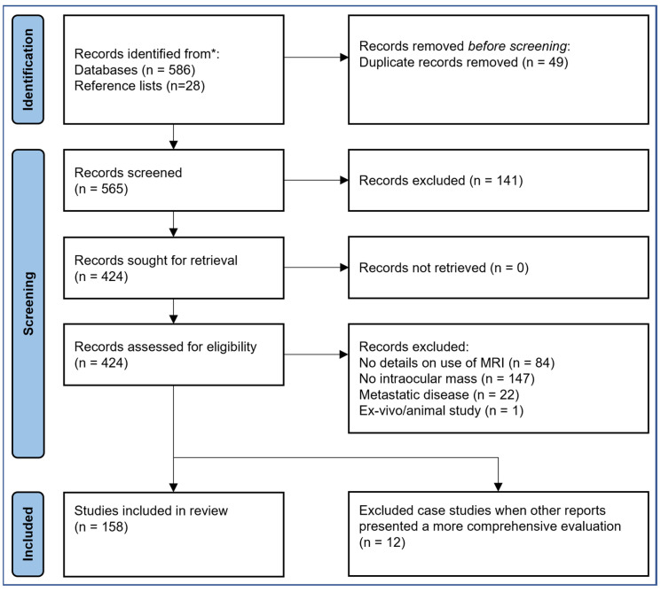
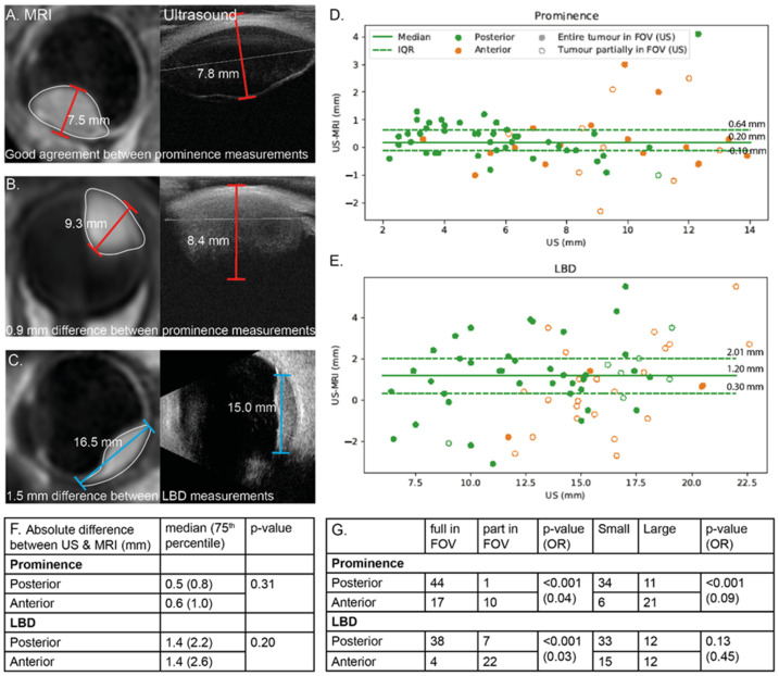
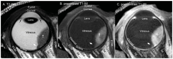
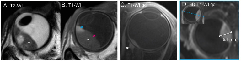
Conversely to most tumour types, magnetic resonance imaging (MRI) was rarely used for eye tumours. As recent technical advances have increased ocular MRI's diagnostic value, various clinical applications have been proposed. This systematic review provides an overview of the current status of MRI in the clinical care of uveal melanoma (UM) patients, the most common eye tumour in adults. In total, 158 articles were included. Two- and three-dimensional anatomical scans and functional scans, which assess the tumour micro-biology, can be obtained in routine clinical setting. The radiological characteristics of the most common intra-ocular masses have been described extensively, enabling MRI to contribute to diagnoses. Additionally, MRI's ability to non-invasively probe the tissue's biological properties enables early detection of therapy response and potentially differentiates between high- and low-risk UM. MRI-based tumour dimensions are generally in agreement with conventional ultrasound (median absolute difference 0.5 mm), but MRI is considered more accurate in a subgroup of anteriorly located tumours. Although multiple studies propose that MRI's 3D tumour visualisation can improve therapy planning, an evaluation of its clinical benefit is lacking. In conclusion, MRI is a complementary imaging modality for UM of which the clinical benefit has been shown by multiple studies.

摘要

与大多数肿瘤类型相反,磁共振成像(MRI)在眼部肿瘤中很少使用。随着近期技术的进步提高了眼部MRI的诊断价值,人们提出了各种临床应用。本系统综述概述了MRI在葡萄膜黑色素瘤(UM)患者临床护理中的现状,UM是成人中最常见的眼部肿瘤。总共纳入了158篇文章。在常规临床环境中可以获得评估肿瘤微生物学的二维和三维解剖扫描以及功能扫描。最常见的眼内肿块的放射学特征已被广泛描述,使MRI有助于诊断。此外,MRI非侵入性探测组织生物学特性的能力能够早期检测治疗反应,并有可能区分高风险和低风险的UM。基于MRI的肿瘤尺寸通常与传统超声一致(中位数绝对差为0.5毫米),但MRI在前部肿瘤亚组中被认为更准确。尽管多项研究表明MRI的三维肿瘤可视化可以改善治疗计划,但缺乏对其临床益处的评估。总之,MRI是UM的一种补充成像方式,多项研究已证明其临床益处。

https://cdn.ncbi.nlm.nih.gov/pmc/blobs/e74b/10252038/a80d39933e05/cancers-15-02995-g006.jpg
https://cdn.ncbi.nlm.nih.gov/pmc/blobs/e74b/10252038/990f7c8569db/cancers-15-02995-g001.jpg
https://cdn.ncbi.nlm.nih.gov/pmc/blobs/e74b/10252038/5b297033ebfd/cancers-15-02995-g002.jpg
https://cdn.ncbi.nlm.nih.gov/pmc/blobs/e74b/10252038/184ac906695d/cancers-15-02995-g003.jpg
https://cdn.ncbi.nlm.nih.gov/pmc/blobs/e74b/10252038/81015ab7b1b4/cancers-15-02995-g004.jpg
https://cdn.ncbi.nlm.nih.gov/pmc/blobs/e74b/10252038/e35797c94de7/cancers-15-02995-g005.jpg
https://cdn.ncbi.nlm.nih.gov/pmc/blobs/e74b/10252038/a80d39933e05/cancers-15-02995-g006.jpg

相似文献

1
Magnetic Resonance Imaging in the Clinical Care for Uveal Melanoma Patients-A Systematic Review from an Ophthalmic Perspective.从眼科视角对葡萄膜黑色素瘤患者临床护理中磁共振成像的系统评价
Cancers (Basel). 2023 May 30;15(11):2995. doi: 10.3390/cancers15112995.
2
Comparison of Magnetic Resonance Imaging-Based and Conventional Measurements for Proton Beam Therapy of Uveal Melanoma.基于磁共振成像的与常规测量在葡萄膜黑色素瘤质子束治疗中的比较。
Ophthalmol Retina. 2023 Feb;7(2):178-188. doi: 10.1016/j.oret.2022.06.019. Epub 2022 Jul 13.
3
The Economic Value of MR-Imaging for Uveal Melanoma.磁共振成像对葡萄膜黑色素瘤的经济价值
Clin Ophthalmol. 2020 Apr 28;14:1135-1143. doi: 10.2147/OPTH.S238405. eCollection 2020.
4
Automatic Three-Dimensional Magnetic Resonance-based measurements of tumour prominence and basal diameter for treatment planning of uveal melanoma.基于自动三维磁共振成像测量葡萄膜黑色素瘤的肿瘤突出度和基底直径以用于治疗规划。
Phys Imaging Radiat Oncol. 2022 Nov 7;24:102-110. doi: 10.1016/j.phro.2022.11.001. eCollection 2022 Oct.
5
Clinical evaluation of ultra-high-field MRI for three-dimensional visualisation of tumour size in uveal melanoma patients, with direct relevance to treatment planning.超高场磁共振成像用于葡萄膜黑色素瘤患者肿瘤大小三维可视化的临床评估,与治疗规划直接相关。
MAGMA. 2016 Jun;29(3):571-7. doi: 10.1007/s10334-016-0529-4. Epub 2016 Feb 25.
6
MRI of Uveal Melanoma.葡萄膜黑色素瘤的磁共振成像
Cancers (Basel). 2019 Mar 17;11(3):377. doi: 10.3390/cancers11030377.
7
MRI and FUNDUS image fusion for improved ocular biometry in Ocular Proton Therapy.MRI 和眼底图像融合在眼部质子治疗中改善眼部生物测量。
Radiother Oncol. 2022 Sep;174:16-22. doi: 10.1016/j.radonc.2022.06.021. Epub 2022 Jul 3.
8
MR imaging characteristics of uveal melanoma with histopathological validation.伴有组织病理学验证的葡萄膜黑色素瘤的磁共振成像特征。
Neuroradiology. 2022 Jan;64(1):171-184. doi: 10.1007/s00234-021-02825-5. Epub 2021 Oct 31.
9
MRI enables accurate diagnosis and follow-up in uveal melanoma patients after vitrectomy.MRI 可在眼葡萄膜黑色素瘤患者玻璃体切除术后进行准确诊断和随访。
Melanoma Res. 2019 Dec;29(6):655-659. doi: 10.1097/CMR.0000000000000568.
10
A Comparison of 3 T and 7 T MRI for the Clinical Evaluation of Uveal Melanoma.3T 和 7T MRI 用于葡萄膜黑色素瘤临床评估的比较。
J Magn Reson Imaging. 2022 May;55(5):1504-1515. doi: 10.1002/jmri.27939. Epub 2021 Oct 15.

引用本文的文献

1
Evaluation of MRI Safety of Ru-106 Eye Applicators.Ru-106 眼部施源器的磁共振成像安全性评估
Ocul Oncol Pathol. 2025 Apr;11(1):13-20. doi: 10.1159/000542712. Epub 2024 Nov 30.
2
Estimating uveal melanoma volume with ellipsoid tumour models.使用椭球体肿瘤模型估计葡萄膜黑色素瘤体积。
Acta Ophthalmol. 2025 Sep;103(6):691-698. doi: 10.1111/aos.17492. Epub 2025 Apr 3.
3
Measurement of Melanocytic Choroidal Lesions: Ultrasound Versus Ultrawide-Field Fundus Imaging System.脉络膜黑素细胞性病变的测量:超声与超广角眼底成像系统对比

本文引用的文献

1
MR-based follow-up after brachytherapy and proton beam therapy in uveal melanoma.磁共振随访在眼黑色素瘤近距离放疗和质子束放疗后的应用。
Neuroradiology. 2023 Aug;65(8):1271-1285. doi: 10.1007/s00234-023-03166-1. Epub 2023 May 30.
2
Automatic Three-Dimensional Magnetic Resonance-based measurements of tumour prominence and basal diameter for treatment planning of uveal melanoma.基于自动三维磁共振成像测量葡萄膜黑色素瘤的肿瘤突出度和基底直径以用于治疗规划。
Phys Imaging Radiat Oncol. 2022 Nov 7;24:102-110. doi: 10.1016/j.phro.2022.11.001. eCollection 2022 Oct.
3
Magnetic resonance imaging reveals possible cause of diplopia after Baerveldt glaucoma implantation.
Cancers (Basel). 2025 Feb 14;17(4):642. doi: 10.3390/cancers17040642.
4
Patient-specific mapping of fundus photographs to three-dimensional ocular imaging.将眼底照片映射到三维眼部成像的个性化方法。
Med Phys. 2025 Apr;52(4):2330-2339. doi: 10.1002/mp.17576. Epub 2024 Dec 12.
5
Quantitative Perfusion-Weighted Magnetic Resonance Imaging in Uveal Melanoma.定量灌注加权磁共振成像在葡萄膜黑色素瘤中的应用。
Invest Ophthalmol Vis Sci. 2024 Sep 3;65(11):17. doi: 10.1167/iovs.65.11.17.
6
Uveal Melanoma: Comprehensive Review of Its Pathophysiology, Diagnosis, Treatment, and Future Perspectives.葡萄膜黑色素瘤:其病理生理学、诊断、治疗及未来展望的综合综述
Biomedicines. 2024 Aug 5;12(8):1758. doi: 10.3390/biomedicines12081758.
7
Geometrical accuracy of magnetic resonance imaging for ocular proton therapy planning.用于眼部质子治疗计划的磁共振成像的几何准确性。
Phys Imaging Radiat Oncol. 2024 Jun 12;31:100598. doi: 10.1016/j.phro.2024.100598. eCollection 2024 Jul.
8
Quantitative Diffusion-Weighted MR Imaging: Is There a Prognostic Role in Noninvasively Predicting the Histopathologic Type of Uveal Melanomas?定量扩散加权磁共振成像:在无创预测葡萄膜黑色素瘤的组织病理学类型方面是否具有预后作用?
Cancers (Basel). 2023 Nov 29;15(23):5627. doi: 10.3390/cancers15235627.
磁共振成像显示 Baerveldt 青光眼植入术后复视的可能原因。
PLoS One. 2022 Oct 20;17(10):e0276527. doi: 10.1371/journal.pone.0276527. eCollection 2022.
4
Commissioning and validation of a novel commercial TPS for ocular proton therapy.一种新型眼部质子治疗商业治疗计划系统的调试与验证
Med Phys. 2023 Jan;50(1):365-379. doi: 10.1002/mp.16006. Epub 2022 Oct 23.
5
Letter to the Editor of Radiotherapy and Oncology regarding the paper titled "MRI and FUNDUS image fusion for improved ocular biometry in Ocular Proton Therapy" by Via et al.致《放射治疗与肿瘤学》编辑的信,关于维亚等人所著题为《用于改善眼部质子治疗中眼生物测量的MRI与眼底图像融合》的论文
Radiother Oncol. 2022 Nov;176:251. doi: 10.1016/j.radonc.2022.08.018. Epub 2022 Aug 18.
6
A descriptive neuroimaging study of retinoblastoma in children: magnetic resonance imaging features.儿童视网膜母细胞瘤的描述性神经影像学研究:磁共振成像特征
Pol J Radiol. 2022 Jul 4;87:e363-e368. doi: 10.5114/pjr.2022.118107. eCollection 2022.
7
Stereotactic irradiation on linear accelerator - ultrasound versus MRI in choroidal melanoma volume calculation.直线加速器立体定向放疗-超声与 MRI 在脉络膜黑色素瘤瘤体计算中的比较。
BMC Ophthalmol. 2022 Aug 5;22(1):333. doi: 10.1186/s12886-022-02558-w.
8
Correlation between clinical presentations, radiological findings and high risk histopathological features of primary enucleated eyes with advanced retinoblastoma at Queen Sirikit National Institute of Child Health: 5 years result.临床表型、影像学发现与高级别视网膜母细胞瘤行眼球摘除术眼高危组织病理学特征的相关性:5 年结果。
PLoS One. 2022 Jul 20;17(7):e0270362. doi: 10.1371/journal.pone.0270362. eCollection 2022.
9
Comparison of Magnetic Resonance Imaging-Based and Conventional Measurements for Proton Beam Therapy of Uveal Melanoma.基于磁共振成像的与常规测量在葡萄膜黑色素瘤质子束治疗中的比较。
Ophthalmol Retina. 2023 Feb;7(2):178-188. doi: 10.1016/j.oret.2022.06.019. Epub 2022 Jul 13.
10
Imaging of Uveal Melanoma-Current Standard and Methods in Development.葡萄膜黑色素瘤的影像学——当前标准及正在研发的方法
Cancers (Basel). 2022 Jun 27;14(13):3147. doi: 10.3390/cancers14133147.